


 Author Affiliations
Abstract
Work-related gun violence is a growing public health concern. Though discussing policy solutions is beyond the scope of this article, one of the more pertinent threats to surviving victims is the retention of lead-containing bullets/bullet fragments. This is a case report of a female of childbearing age with retained lead bullet fragments from a work-related gunshot wound to the spine that traversed the pelvis and caused symptomatic elevated blood lead levels. The patient presented for a pre-pregnancy consultation due to retained lead bullet fragments with Occupational and Environmental Medicine. She had experienced elevated blood lead levels many months after the gunshot wound. This case discusses the difficulty with interpreting lead guidance in females attempting pregnancy planning, particularly with retrained lead fragments, limited surgical options in gunshot wound survivors, and challenges with workers’ compensation coverage for female fertility and potential chronic impairments in offspring. Recommendations are for greater workplace violence and firearm protections, comprehensive lead guidance for pregnancy planning with diverse lead exposures, and workers’ compensation coverage of females protecting fertility and offspring health affected as a result of work-related injuries. The authors summarize existing relevant guidelines and briefly outline an algorithm for clinicians caring for such patients. Though political prevention is unlikely to occur soon, clinical management has significant potential to reduce suffering and long-term sequelae.
Keywords
Gunshot wound, Retained lead fragments, Pre-pregnancy planning, Lead toxicity, Workers’ compensation.
Learning Points
What is already known about this subject:
- Gun violence is one of the leading causes of occupational fatalities
- Retained lead bullet fragments from gunshot wounds can cause clinically significant elevations in blood lead levels
- Chronic lead exposure is neurotoxic in adults, and developing fetuses are especially vulnerable
What this case report adds:
- A clinical assessment of a female of childbearing age with acute and chronic injuries from workplace gun violence, that resulted in retained lead bullet fragments, symptomatically elevated blood lead levels, pelvic injuries, and long-term impairments, who desires pregnancy
- Clinical evidence demonstrating devastation of workplace gun violence and chronic lead exposures to female survivors of childbearing age, with subsequent risk to their fertility and potential future health outcomes of offspring
What impact does this may have on practice, policy, or procedure?
- Recognition of complexity and compounding risks to female fertility and offspring from workplace gun violence and chronic lead exposure
- Need for workers’ compensation to support coverage to include protection of women’s fertility and anticipation of adverse pregnancy complications and/or health outcomes in offspring from work-related injuries or illnesses due to gun violence and lead exposures
- Updated work protections, standards, and research with consistent and optimized guidance for women of childbearing age with diverse types of lead exposures seeking pregnancy
Introduction
Work-related gun violence is a growing public health concern that is associated with significant mortality and morbidity.[1] According to the Centers for Disease Control (CDC) and Prevention data from 2021-2022, the United States had 48,204 firearm-related fatalities, accounting for 21.7% of total deaths.[2] Workplace violence is among the leading causes of fatal occupational injuries after transportation incidents and falls.[3,4] Nearly 70% of firearm-related injuries are nonfatal, and retained lead fragments are an important cause of lead toxicity appearing years later.[1] Retained lead bullet fragments from a gunshot wound may cause long-term adverse health effects due to the physical locations or toxicity from lead.[1] Instances of retained lead fragments are rarely reported in the literature, and there is a scarcity of guidance for women planning pregnancy with retained fragments and lead toxicity. Lead is a heavy metal with unique properties that can disrupt an array of cellular mechanisms throughout the body.[5] Lead poisoning is typically gradual and asymptomatic, aside from the elevated lead concentration itself. Commonly, the onset of lead toxicity symptoms is non-specific but may include fatigue, paresthesia in hands or feet, hypertension, memory difficulties, headaches, insomnia, or abdominal pain.[6] A high index of suspicion is needed to prompt a test for lead in patients with retained bullet fragments.
We present a rare case of a female of childbearing age actively attempting pregnancy after a work-related gunshot wound with retained lead bullet fragments. The patient has elevated lead levels, fragments posing a mechanical childbirth risk, impaired fertility risk, risk of pregnancy-related complications, and risk of adverse fetal neurodevelopmental outcomes. Work-related gun violence injuries and elevated blood lead levels affecting women of childbearing age represent compounded risk in workers’ compensation cases due to long-term effects, lost time, disability, impact on fertility, and risk to offspring.
Case Presentation & Management
A 33-year-old primiparous female was referred by her primary care clinician to Occupational and Environmental Medicine (OEM) due to concern regarding elevated blood lead levels from a gunshot wound (GSW) sustained sixteen months prior while at work. A gunman shot the employee in her lower back while at work in a healthcare facility, resulting in numerous retained lead fragments in the lumbar spine and two large bullet fragments in the pelvis. The patient presented recommendations seeking pre-pregnancy counseling. Initial injuries from the gunshot wound included an L5 spinous process fracture, L5-S1 comminuted fractures, rectal tear, right fallopian tube and ovary injury. Acute care included a two-week hospital admission with surgical exploratory laparotomy, small bowel resection, and repair of the rectal tear. Her post-hospital course covered multidisciplinary management with neurosurgery, trauma surgery, urology, primary care, psychology, and physical therapy.
The patient’s primary care provider ordered venous lead testing due to bilateral hand and finger tingling, which revealed elevated lead levels of 17 ug/dL and 15 ug/dL at 7 months and 11 months post-GSW, respectively. Imaging included pelvic and lumbar spine X-rays and CT at 7.5 months post-injury that demonstrated the largest bullet fragment lodged in the right perirectal fat and another fragment in the obturator foramen, many metallic foreign bodies in chronic comminuted fractures of L5-S1, and a 3.2 cm cystic collection in the right anterior sacrum abutting the thecal sac (see Figures 1 and 2).
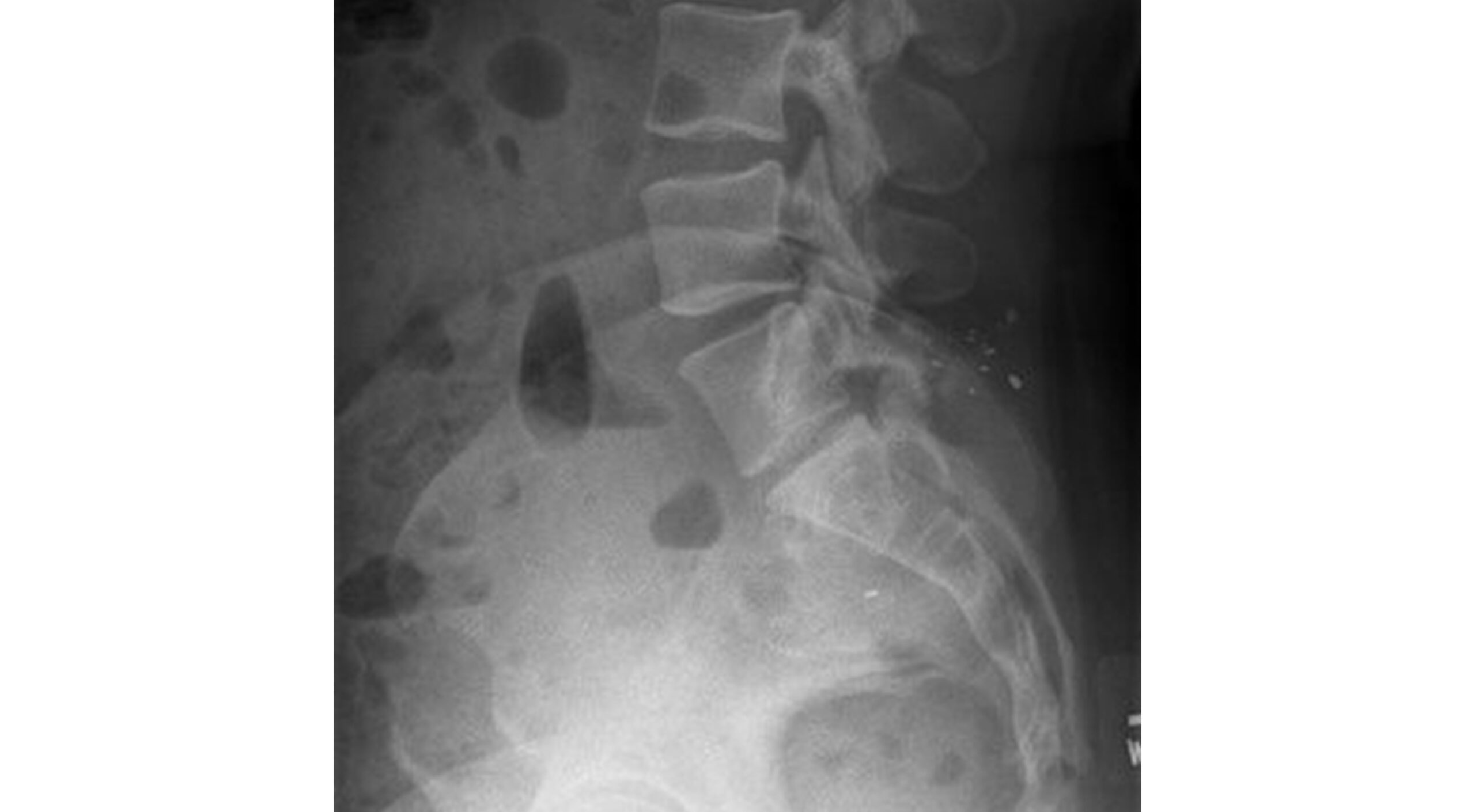
Figure 1: Lateral Lumbar Spine X-ray demonstrating lead bullet fragments in the posterior L5- S1 vertebra. As well as L5 spinous process fracture, and L5-S1 comminuted fractures. Cystic collection with multiple lead fragments in the right anterior sacrum
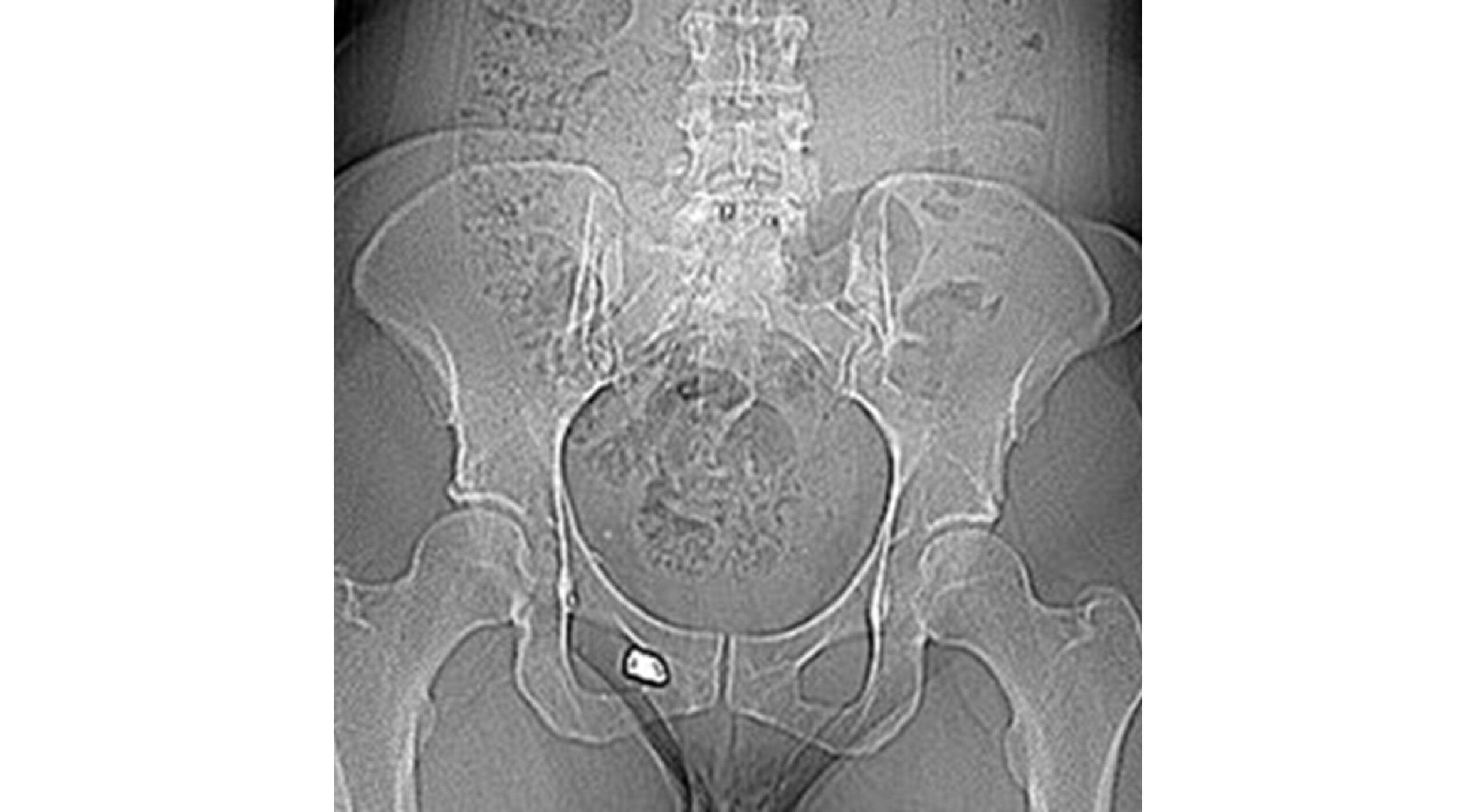
Figure 2: AP Pelvis X-ray demonstrating the main bullet fragment of the right obturator foramen. Lead fragments in L5-S1. Constipation and lead fragments in a cystic collection abutting the right anterior sacrum
At 10 months post-GSW, maternal-fetal medicine (MFM) was consulted because the patient was actively attempting pregnancy with significant anatomic concerns due to the bullet traversing her spine and pelvis and elevated lead levels. MFM recommendations included American College of Obstetrics and Gynecology (ACOG) guidance for periodic lead monitoring up to and at delivery for venous lead levels of 14-25 µg/dL7, consideration of alternative lead sources, and ultrasound monitoring for ectopic pregnancy. Primary care referred the employee to the Environmental Medicine clinic for consultation.
Sixteen months post-GSW, she presented to the OEM clinic, and her medical impairments involved bilateral hand paresthesia, unilateral lower extremity paresthesia and weakness, neurogenic bladder requiring self-catheterization, fecal incontinence, dizziness, post-traumatic stress disorder, and chronic low back pain. Due to the ongoing severity of the employee’s impairments and her rehabilitation, she has not returned to work. Exposure history demonstrated no additional lead exposures.
Upon thorough review of medical records, the consultation focused on lead and risk reduction. Recommendations included repeating venous lead levels every 3 months and referral for a second surgical opinion. The employee was referred to a different surgical obstetrician for re-evaluation of any mechanical risks or potential removal of lead fragments. Multidisciplinary surgical consultation, including neurosurgery and colorectal surgery, yielded too many risks for further surgical intervention. Follow-up lead testing revealed levels of 13 and 12 ug/dL at 15 and 17 months post-GSW, respectively. The patient has still not become pregnant.
Discussion
Chronic lead exposure in adults may reduce cognitive performance, whereas in fetuses or infants who are more sensitive, it is associated with behavioral problems, learning deficits, and lowered intelligence quotients (IQ).[6] Treatment for elevated lead levels includes removing sources of exposure; however, surgical removal is often untenable. In this case, various surgeons explained the difficulty of removal due to extensive scar tissue and the difficulty of reaching the anatomical location abutting the bowel wall, neurovascular supply, or thecal sac with a greater risk of causing further harm. Optimizing the nutritional status of calcium, iron, folate, and zinc intake is highly recommended due to these nutrients potentially displacing lead when orally ingested. At markedly elevated lead concentrations, a variety of chelating agents may be considered alone or in combination. However, chelating agents can be potentially harmful, depleting other nutrients and mobilizing lead storage from bones, without typically reversing cognitive deficits.[6]
Diagnostic testing often begins with a complete blood count (lead poisoning may be associated with iron deficiency anemia), blood film/smear (showing basophilic stippling). Lead poisoning may be associated with increased erythrocyte protoporphyrin (EPP), which increases when lead in the blood is high or in iron deficiency, with a delay of several weeks. Measurement of blood lead levels is associated with recent or current lead exposure and does not give an accurate estimation of lead stored primarily in the bones. The latter is particularly important in pregnant patients, where bone stores may become mobilized starting in the first trimester during intensive fetal development and during lactation.[7] Lead storage in bones or total lead body burden may be measured by X-ray fluorescence.[6] Blood lead levels appear to be declining in this patient, which may be explained by lead storage in bones, but may increase when mobilized during pregnancy and lactation. Lead exposure, acute or cumulative, during pregnancy is associated with increased risk of gestational hypertension, spontaneous abortion, low birth weight, and impaired fetal neurodevelopment.[7] Currently, there is uncertainty regarding maternal lead levels and their association with the magnitude of increasing risk for pregnancy-related complications or adverse fetal outcomes.[7,8] Lead can be transmitted from mother to baby via breast milk; therefore, babies should be monitored.[9] In essence, there is no safe lead level for pregnancy or in children. Lead levels below 10 ug/dL still demonstrate inverse relationships with fetal growth/neurodevelopment.[8]
Ambiguity and concerns persist with Clinical guidance on the management of lead in pregnancy. The CDC recommends blood lead levels below five ug/dL during pregnancy and above this, periodic monitoring; however, this guidance does not take into consideration total body lead burdens that may become mobilized from bones.[7,8] Pediatric lead levels above 3.5 ug/dL are considered hazardous based on recent CDC guidance.[10] The inconsistent guidance for lead levels for pregnant women and for children further does not address whether chelation would be practical at such low doses or create greater risks.
Lead exposures in women and pregnancy raise more challenges to workers’ compensation insurers and concerns over coverage if female fertility is at risk or when reproductive capacity is maintained with risks to the health of the mother and fetus arise. There are occupations where women have lead exposures and periodic blood lead level monitoring may not be enough to capture total body burden or risk to pregnancy and offspring.[11] The Occupational Safety and Health Administration (OSHA) 1978 Lead standard has removal level only if whole blood levels are above 60 μg/dL in general industry, and for women planning to become pregnant, lead levels of under 30 μg/dL.[12] This case demonstrates the shortfalls of workers’ compensation coverage and the need for reform of OSHA lead standards. Medicolegally, there are arguments that dismiss coverage for the health outcomes of a child as the child was not an employee, whereas a counterargument could include the fact that the child’s adverse health outcomes would not have occurred but for the work-related injury. This may be further complicated if the child is found to have long-term adverse cognitive effects that may require lifelong care beyond the neonatal period.
At present, such clinical scenarios of lead contamination in pregnant workers represent a small patient population, with guidance coming either from individual researchers13 or adjacent medical societies.[7-9,11,12,14]. While other case studies merely outline the dramatic impact of the bullet or other exposure, without offering guidance.[15] In light of this lack of centralized guidance, the authors would like to briefly describe, in this section, each guiding body’s recommendations and offer a unified algorithm. The American College of Obstetrics and Gynecology (ACOG) does not recommend routine screening for lead during pregnancy, though risk assessment should be monitored closely. Exposure control and counseling are recommended above 5 ug/dL, and breastfeeding should be stopped above 40 ug/dL until it is brought down below this level, with expert consultation sought at 45 ug/dL.[14]
In the work setting, OSHA advises in 1910.1025(k)(1)(i)(B) that should the employee be removed, “The employer shall remove an employee from work having an exposure to lead at or above the action level on each occasion that the average of the last three blood sampling tests conducted pursuant to this section (or the average of all blood sampling tests conducted over the previous six (6) months, whichever is longer) indicates that the employee’s blood lead level is at or above 50 µg/100 g of whole blood; provided, however, that an employee need not be removed if the last blood sampling test indicates a blood lead level below 40 µg/100 g of whole blood.”[11]
The CDC based its guidance of 3.5 ug/dL due to it representing the 97.5th percentile (or 2 standard deviations) above the mean for US children ages 1-5 years old.[12] While they do recommend universal screening for lead at ages 12 and 24 months old, regarding pregnant women, they advise increased monitoring (with possible environmental health sampling or questionnaires like those developed by the New York City Department of Health and Mental Hygiene or the Minnesota Department of Health) above 5 ug/dL and below 39 ug/dL.[8] They further emphasize that if the mother’s BLL is greater than 20 ug/dL and the infant’s BLL is greater than 5 ug/dL with suspected exposures controlled, it likely points to the breast milk as being the source of exposure. ACOG adopted their recommendation to cease breastfeeding above 40 ug/dL from the CDC, which also advises pumping and discarding the high lead breast milk for the mother’s safety. Universally, the CDC also advises not to drink tap water with greater than 15 parts per billion lead.
Having outlined guidance above, the authors recognize that not all that is advised is directly applicable in a clinical setting and consider when issuing these suggestions for intervention. When facing a lead-exposed pregnant patient, universal blood lead level screening is not recommended unless a risk factor is identified. Patient counseling and exposure control with the Department of Health sampling is appropriate above 3.5 ug/dL. Breastfeeding can be continued below 40 ug/dL, which is also the threshold for removal from the workplace, if the exposure is suspected to be occupational. Individuals with greater than 45 ug/dL should seek toxicology, occupational medicine, or other expert consultation and defer to them for chelation guidelines.
Conclusion
Management of work-related gun violence and lead toxicity in females is a complex case due to long-term health impairments, fertility risk, pregnancy-related complications, and risk to the fetus with limited medical or surgical options. Workers’ compensation coverage is unclear regarding accounting for a woman’s reproductive capacity or adverse health outcomes in offspring that may result from a work-related injury. Worker protections and standards need to evolve to protect females with reproductive capacity and children. This case raises significant practical concerns for workers’ compensation in terms of preventable costs and adverse health outcomes that could be mitigated with changes in public policy related to workplace gun violence and revised lead standards.
References
- Weiss D, Tomasallo CD, Meiman JG, et al. Elevated Blood Lead Levels Associated with Retained Bullet Fragments – United States, 2003-2012. MMWR Morb Mortal Wkly Rep. 2017;66(5):130-133. doi:10.15585/mmwr.mm6605a2
PubMed | Crossref | Google Scholar - Centers for Disease Control and Prevention. Multiple Cause of Death Data on CDC WONDER. 2025.
Multiple Cause of Death Data on CDC WONDER - U.S. Bureau of Labor Statistics. Census of Fatal Occupational Injuries — 2023. USDL-24-2564; 2024.
Census of Fatal Occupational Injuries — 2023 - U.S. Bureau of Labor Statistics. Injuries, Illnesses, and Fatalities (IIF) program. 2022.
Injuries, Illnesses, and Fatalities (IIF) program - Wani AL, Ara A, Usmani JA. Lead toxicity: A review. Interdiscip Toxicol. 2015;8(2):55-64. doi:10.1515/intox-2015-0009
PubMed | Crossref | Google Scholar - Rubin R. Rubin’s Pathology: Clinicopathologic Foundations of Medicine. 6th ed. Jefferson Faculty Books; 2012.
Rubin’s pathology: clinicopathologic foundations of medicine - Committee on Obstetric Practice. Committee opinion No. 533: lead screening during pregnancy and lactation. Obstet Gynecol. 2012;120(2 Pt 1):416-420. doi:10.1097/AOG.0b013e31826804e8
PubMed | Crossref | Google Scholar - CDC. Guidelines for the Identification and Management of Lead Exposure in Pregnant and Lactating Women. 2021. Guidelines for the identification and management of lead exposure in pregnant and lactating women
- CDC. Lead and Breastfeeding. 2025.
Lead and Breastfeeding - CDC. CDC Updates Blood Lead Reference Value. 2024.
CDC Updates Blood Lead Reference Value - La-Llave-León O, Salas Pacheco JM, Estrada Martínez S, et al. The relationship between blood lead levels and occupational exposure in a pregnant population. BMC Public Health. 2016;16:1231. doi:10.1186/s12889-016-3902-3
Crossref | Google Scholar - U.S. Department of Labor, Occupational Safety and Health Administration. Regulations (Standards – 29 CFR).
Regulations (Standards – 29 CFR) - Apte A, Bradford K, Dente C, Smith RN. Lead toxicity from retained bullet fragments: A systematic review and meta-analysis. J Trauma Acute Care Surg. 2019;87(3):707-716. doi:10.1097/TA.0000000000002287
PubMed | Crossref | Google Scholar - American College of Obstetrics & Gynecology (ACOG). Lead Screening During Pregnancy and Lactation. 2012; Number 533.
Lead Screening During Pregnancy and Lactation - Raymond LW, Ford MD, Porter WG, Saxe JS, Ullrich CG. Maternal-fetal lead poisoning from a 15-year-old bullet. J Matern Fetal Neonatal Med. 2002;11(1):63-66. doi:10.1080/jmf.11.1.63.66
PubMed | Crossref | Google Scholar
Acknowledgments
No further acknowledgements are warranted.
Funding
None
Author Information
Corresponding Author:
Dominik Dabrowski
Department of Occupational and Environmental Medicine, HealthPartners, St. Paul, USA
School of Public Health, University of Minnesota, Minneapolis, USA
Email: dabrowskidom@gmail.com
Co-Authors:
Ashley Nadeau, Zeke J. McKinney
Department of Occupational and Environmental Medicine, HealthPartners, St. Paul, USA
School of Public Health, University of Minnesota, Minneapolis, USA
Beth Baker
School of Public Health, University of Minnesota, Minneapolis, USA
Authors Contributions
All authors had full access to the data and actively participated in the development of the manuscript. AN contributed to the conceptualization of the study, acquisition of data, and preparation of both the original draft and subsequent revisions. DD was involved in data acquisition, visualization, drafting the original manuscript, and reviewing and editing the final version. ZJM and BB provided supervision, contributed to the study’s conceptualization, and participated in reviewing and editing the manuscript.
Informed Consent
The patient provided fully informed consent and agreed to the use of their case information, provided it was completely anonymized.
Conflict of Interest Statement
The authors declared no conflict of interest.
Guarantor
None
DOI
Cite this Article
Nadeau A, Dabrowski D, Baker B, McKinney ZJ. Retained Lead Bullet Fragments in Woman of Childbearing Age. medtigo J Emerg Med. 2025;2(4):e3092245. doi:10.63096/medtigo3092245 Crossref


