


 Author Affiliations
Abstract
Introduction: Pathology specimens constitute a cornerstone of undergraduate medical education, serving as an essential bridge between theoretical knowledge and clinical application. Exposure to preserved human organ specimens enables students to visualize disease processes, understand morphological changes, and correlate pathological findings with clinical manifestations, thereby strengthening diagnostic reasoning and critical thinking skills.
Objective: To evaluate medical students’ perceptions, opinions, and experiences regarding the effectiveness of pathology specimens in learning at the Department of Pathology, Fazaia Medical College, Air University, Islamabad.
Methodology: A cross-sectional descriptive study was conducted using an online survey among third- and fourth-year undergraduate MBBS students (n = 90). Data were collected using a pre-tested questionnaire and analyzed using the Statistical Package for the Social Sciences (SPSS) software. Descriptive statistics were applied, and the Chi-square test was used to assess associations among categorical variables.
Results: A majority of students (82%) perceived pathology specimens as an effective teaching–learning tool. Additionally, 76% agreed that hands-on experience enhanced their understanding, and 66% felt adequately prepared to apply their learned knowledge in clinical settings. Most participants preferred physical pathology museums over virtual alternatives, underscoring the importance of tangible learning experiences.
Conclusion: Pathology museums containing preserved human organ specimens significantly enhance medical students’ learning and critical thinking skills.
Keywords
Medical education, Pathology specimens, Learning, Medical students, Clinical reasoning.
Introduction
In medical education, pathology specimens are essential for helping students grasp the structural and functional changes that arise in various disease processes. Pathology education focuses on expanding medical students’ knowledge of disease mechanisms. Traditionally, medical schools have taught pathology through a blend of theoretical lectures, practical activities, clinical autopsies, and the exhibition of micro and macroscopic specimens in pathology museums.[1] Through hands-on interaction with pathology specimens/ biopsies, such as tissue samples, organs, and histological slides, students gain a deeper understanding of disease processes, diagnostic techniques, and treatment modalities. These specimens provide a tangible representation of the pathology being studied, allowing students to correlate clinical manifestations. Since the early 19th century, pathology museums with a collection of different organ pathology specimens have been instrumental in medical education. Pathology specimens are labelled and preserved in formalin-based solutions enclosed in glass jars, which are exhibited in the pathology museums. This setup allows medical students to explore gross specimens and microscopic images and therefore encourages self-directed learning. Small group discussions and practical classes also become possible with the physical layout of these museums. For the pathology museum to sustain peak quality, it should be meticulously arranged, visually enticing, and subject to regular updates, both with the variety of pathological processes affecting the human tissues and their changing diagnosis due to new histopathological techniques.[2]
In clinical practice, medical students need to learn critical thinking and clinical reasoning skills. Studying pathology and using diseased organs from the pathology museum (known as ‘pots’) can help students to learn critical thinking and clinical reasoning skills. This is through the appreciation of the pathological basis of clinical symptoms and signs in patients, reinforcing situations observed during their clinical rotations. In the domain of clinical practice, it is vital for medical students to develop their analytical thinking and clinical decision-making skills. Learning pathology and utilizing specimens from the pathology museum, often referred to as ‘ ‘, can be instrumental in fostering these skills among students. This involves understanding the pathological foundation of clinical symptoms and signs in patients and reinforcing observations made during their clinical rotations.[3]
Students learn pathology material at varying rates, and when they enter pathology residency, they have differing levels of prior experience. It is significant to ensure that material or equipment that is unavailable at home is used for on-campus interactive lectures and discussions for the medical students. The example of this for pathology includes physical presence of gross organs, anatomic dissection, and other technical activities of handling the gross and microscopic representative sections, such as frozen sections and cytologic preparations.[4,5]
To assess the perceptions of medical students in Fazaia Medical College for effective utilization of Pathology gross specimens and models for learning pathology. The synopsis was presented to the Institutional Review Board of Fazaia Medical College, and formal approval was sought.
After reviewing the literature, it was revealed that there was no study that has yet been published which has collected data regarding the perceptions and enhancement in learning of Pathology while using human organ Pathology specimens and models. At Fazaia Medical College, Air University, Islamabad, Pakistan, we have put in efforts to regularly use Pathology specimens for teaching, learning, and assessment. We decided to collect the data regarding the above knowledge gap and analyze it.
Methodology
This study was a cross-sectional descriptive study conducted through an online survey for data collection of undergraduate Year 3 and Year 4 students at Fazaia Medical College, Islamabad, at Air University E-9 Campus. Year 1 and Year 2 students were not included in the survey due to their insufficient exposure to pathology at that time, as the curriculum is integrated, and pathology is primarily introduced in Year 3. The study period spanned from June 1, 2024, to June 30, 2024. Out of 120 eligible students in Year 3 and Year 4, 90 students consented to participation, resulting in a response rate of 75%. A convenience sampling method was used to select this study sample, which included all participating students.
Ethical approval was obtained from the Institutional Review Board of Fazaia Medical College, Air University, Islamabad (Reference No: IBD/FMC/134/IRB/5).^4 Informed consent was obtained from all participants prior to their participation, and assurances of confidentiality and voluntary participation were provided.
To ensure reliability, a questionnaire was developed in English and pre-tested with 30 MBBS students, resulting in a Cronbach’s alpha of 0.81. The questionnaire comprised items covering students’ frequency of museum visits, perceived effectiveness of specimens, impact on hands-on learning, preparedness for clinical settings, preference for physical vs. virtual museums, and impact on collaborative learning. An expert review was conducted by two senior pathologists to ensure content validity. The final questionnaire was distributed via Microsoft Forms, and invitations to participate were sent to students using the WhatsApp messaging platform. This approach facilitated easy access and a high response rate from the targeted student population. All submitted data were summarized in Microsoft Excel and analyzed using the statistical package for the social sciences (SPSS) software version 26. A chi-square test was conducted to identify associations within the categorical data, specifically examining the relationship between gender and perceptions of specimen effectiveness. Only respondents who provided written consent to participate were included in the analysis.
Results
A total of 90 undergraduate medical students (Year 3 and Year 4) participated in this study. Of the respondents, 65 (72.2%) were female, and 25 (27.8%) were male. No missing data was observed, as participation in all questionnaire items was mandatory for submission. Categorical data were coded as per standard practice for SPSS analysis.
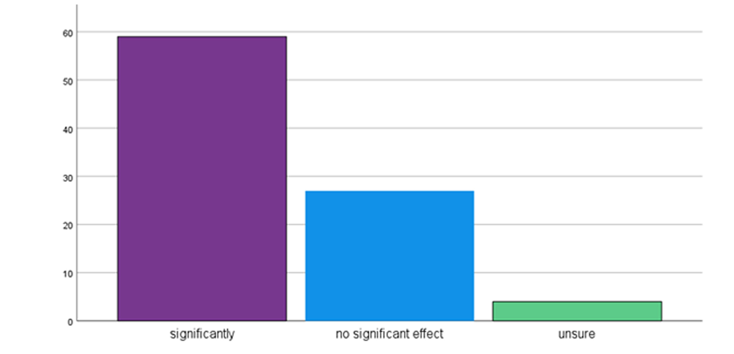
Figure 1: Frequency of pathology museum utilization by undergraduate medical students (N = 90)
Figure 1 illustrates the frequency of students’ visits to the Pathology museum. A substantial proportion of students, 46 (51.1%), reported utilizing the museum on a weekly basis, highlighting regular engagement with the specimens. This was followed by 30 students (33.3%) who visited monthly, 15 students (16.7%) rarely/never, and only four students (4.4%) who visited daily.
Table 1 summarizes students’ perceptions of the effectiveness of pathology specimens in their education. A substantial majority of respondents, 74 students (82.2%), considered pathology specimens to be either “effective” (40 students, 44.4%) or “very effective” (34 students, 37.8%) in their undergraduate medical education. Only five students (5.6%) perceived them as “not effective”, while 11 students (12.2%) were “neither effective nor ineffective.
| Effectiveness category | Number of students | Percentage (%) |
| Not effective | 5 | 5.6 |
| Neither effective nor ineffective | 11 | 12.2 |
| Effective | 40 | 44.4 |
| Very effective | 34 | 37.8 |
| Total | 90 | 100.0 |
Table 1: Perceived effectiveness of pathology specimens (N = 90)
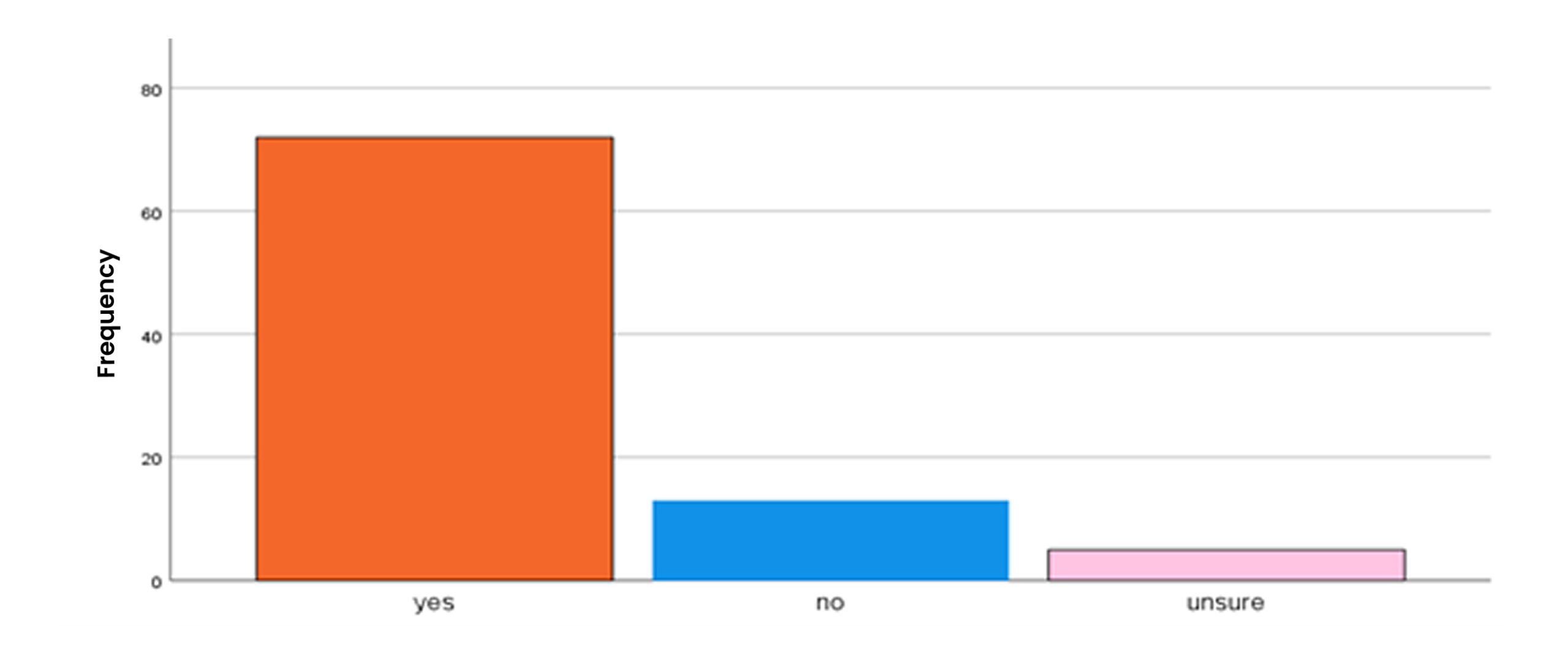
Figure 2: The impact of hands-on experience
Regarding the impact of hands-on experience, 68 students (75.6%) affirmed that hands-on interaction with pathology specimens enhances their learning. In contrast, 15 students (16.7%) stated it did not improve their learning, and seven students (7.8%) were unsure.
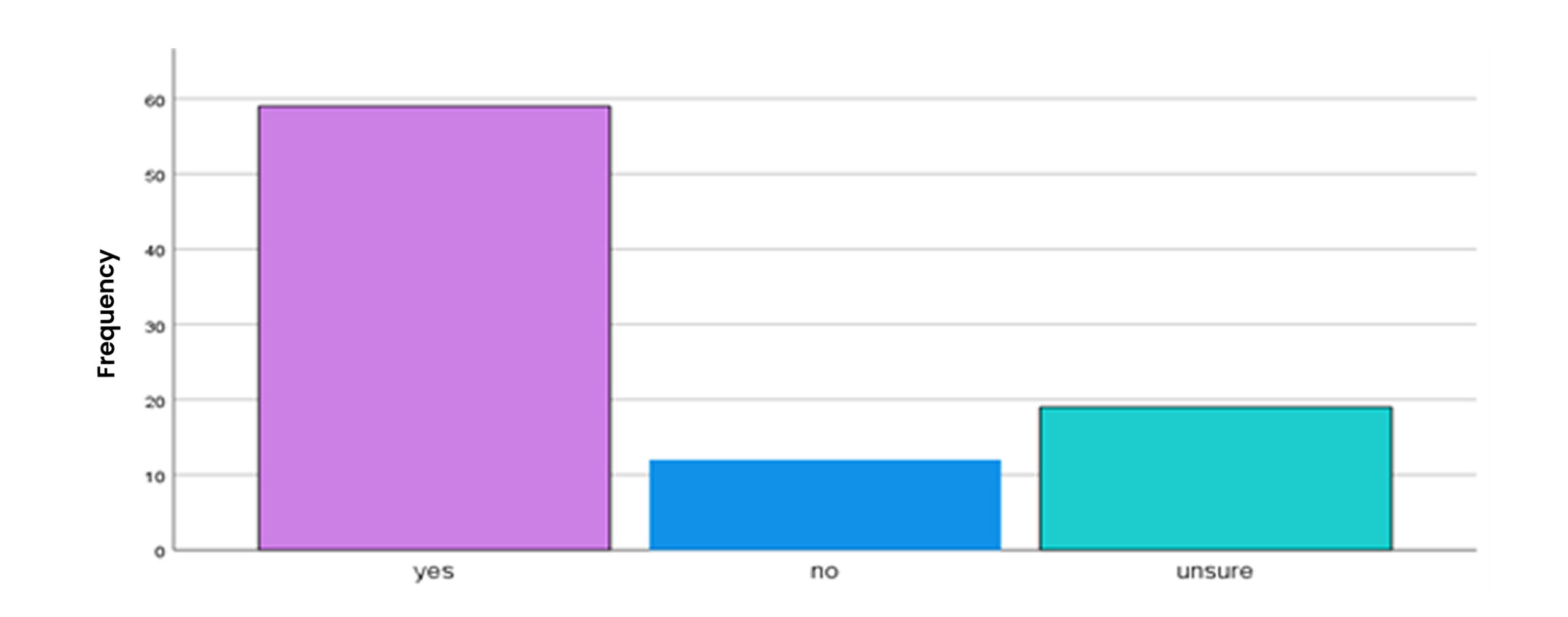
Figure 3: Preparedness for clinical application
Fifty-nine students (65.6%) expressed confidence, indicating that they felt adequately prepared to apply the knowledge acquired from using pathology specimens in practical clinical situations. Conversely, 15 students (16.7%) did not feel prepared, and 16 students (17.8%) were unsure.
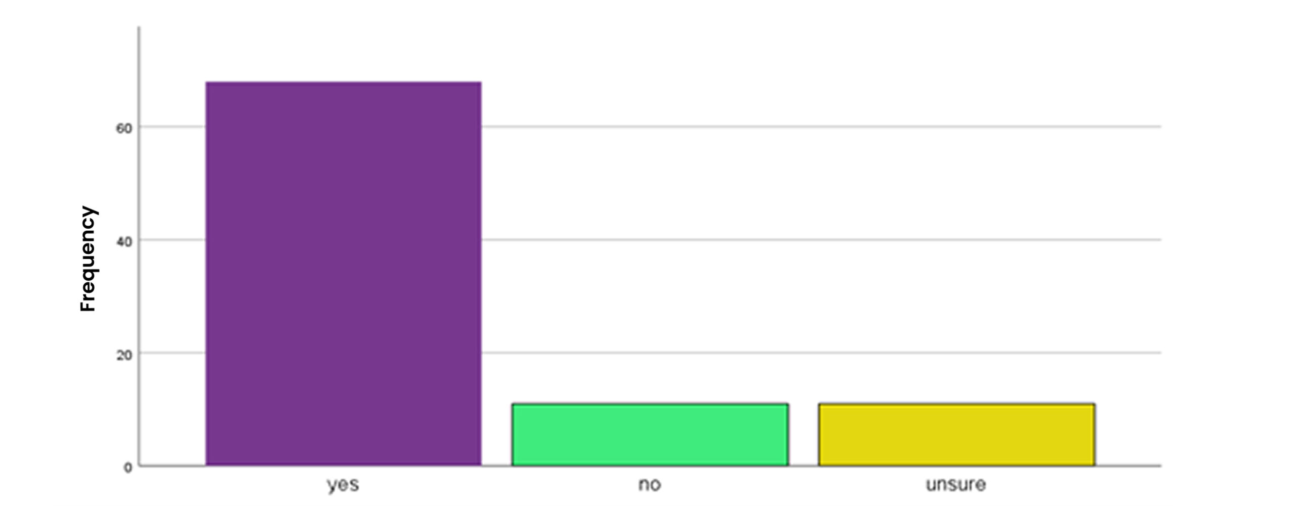
Figure 4: Preference for physical vs. virtual pathology museums
When asked about their preference between physical and virtual pathology museums, 72 students (80%) expressed a clear preference for a physical pathology museum over a virtual one. Only seven students (7.8%) preferred virtual museums, and 11 students (12.2%) were unsure.
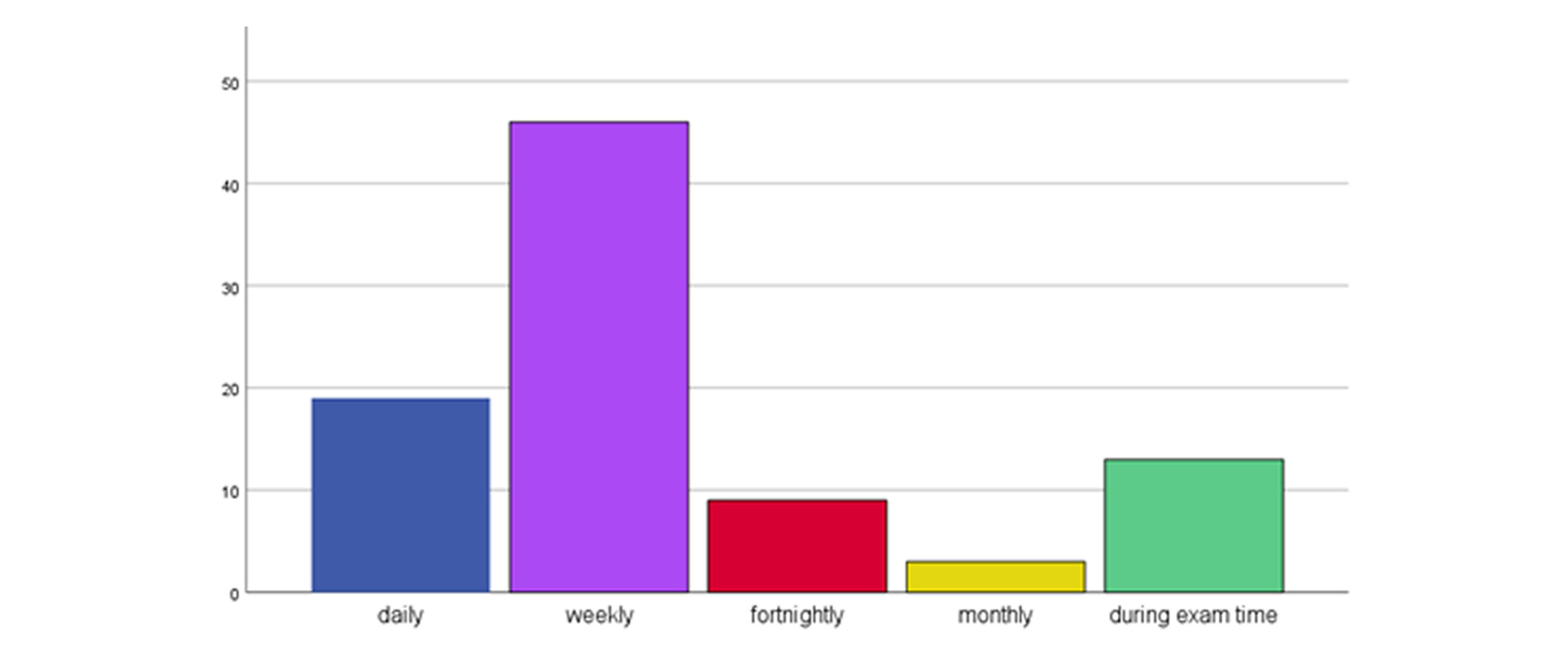
Figure 5: Impact on collaborative learning experiences
The use of pathology specimens had a positive impact on collaborative learning for many students. Fifty-nine students (65.6%) felt that the use of pathological specimens had a substantial positive effect on their collaborative learning experiences. Twenty students (22.2%) reported no significant effect, and 11 students (12.2%) were unsure. Table 2 presents the results of the Chi-square tests conducted to explore associations between key categorical variables.
Most medical students reported visiting the pathology museum because it was mandatory. They were generally satisfied with the museum’s accessibility. The pathology specimens inspired many students to consider a career in pathology and fostered a sense of collaboration among them. Additionally, the majority of students reported receiving feedback on their understanding of the specimens. However, most students felt that the specimens should be complemented with other teaching methods. A Chi-square test was conducted to examine whether there was an association between gender and the perceived effectiveness of pathological specimens.
| Association | Chi-square (χ²) | Degrees of freedom (df) | p-value |
| Gender and Perceived Effectiveness of Specimens | 2.15 | 3 | 0.542 |
| Frequency of Museum Utilization (Weekly vs. Less Frequent) and Preparedness for Clinical Application | 0.98 | 1 | 0.322 |
Table 2: Chi-square test results for associations between key variables
No statistically significant association was found between gender and the perceived effectiveness of pathology specimens (χ² (3, N = 90) = 2.15, p = 0.542), indicating that both male and female students perceived the effectiveness of specimens similarly. Similarly, there was no significant association between the frequency of museum utilization (categorized as weekly vs. less frequent) and feeling prepared for clinical application (χ² (1, N = 90) = 0.98, p = 0.322). This suggests that in this cohort, more frequent museum visits did not statistically correlate with a higher self-reported sense of preparedness for clinical practice.
Discussion
This study revealed that a majority of undergraduate medical students (82%) at Fazaia Medical College perceived pathology specimens as an effective or very effective mode for teaching and learning. This aligns with previous research highlighting the significant role of tangible interactions in medical education. Our findings indicate that 76% of students affirmed that hands-on experience with pathology specimens enhances their learning, a result consistent with a study conducted in Kuala Lumpur, Malaysia, where practical classes were the most preferred way for learning pathology among students.[1] This similarity suggests a universal appreciation among medical students for active, tactile engagement with learning materials, as it provides a direct visual and physical correlation to theoretical knowledge.
The practical aspect of the traditional teaching approach, which involves examining macroscopic specimens and histology slides in medical museums, alongside utilizing clinical autopsies, is strongly supported by our findings.[5] This outcome underscores the enduring value of traditional methods in pathology education. Studies consistently highlight the value of hands-on learning, which allows students to observe and interact with gross and microscopic pathology in a way that traditional lectures or digital resources may not fully replicate.[4,6] These physical interactions stimulate visual and kinaesthetic learning, which reinforces memory and deepens understanding. Research has shown that students engaging with actual specimens demonstrate improved diagnostic skills and a better grasp of complex pathological concepts.[5]
A further significant finding of our study is that 66% of students felt adequately prepared to apply their learned knowledge from specimens in clinical settings. This confidence in clinical application reinforces the direct relevance of specimen-based learning to professional practice. Similar observations were made by Taylor et al.[7] who found that students actively participating in practical classes and utilizing pathology specimens reported feeling 40% more prepared for clinical practice compared to those who did not engage with these resources. This emphasizes that direct exposure to pathological changes in organs and tissues helps students bridge the gap between theoretical knowledge and real-world clinical manifestations, thereby strengthening their diagnostic and reasoning skills.
Moreover, our study found a strong preference among students (80%) for a physical pathology museum over a virtual one, despite the increasing availability of digital resources. This preference suggests that while virtual tools offer flexibility, they may not fully replicate the sensory and contextual learning experience provided by physical specimens. This is crucial as a study by Vaduva et al.[8] Noted a decrease in student performance upon switching to purely digital models, where the mean overall marks of Year-2 medical students increased from 65.36% in 2019 to 75.83% in 2020 after switching to online learning, highlighting an improvement of approximately 10.47%. While this study suggests an improvement with online learning, the overall preference for physical museums in our study and the concerns raised by others about digital-only models indicate that a balanced approach is likely most effective. The integration of both physical and digital resources, where digital tools complement rather than replace physical specimens, could maximize learning outcomes. Such an approach could address challenges like accessibility and provide supplementary information while retaining the benefits of hands-on interaction.
Despite the clear educational benefits, pathology museums face several challenges identified in our study and the literature. These include the need for regular updates and meticulous maintenance of specimens, improving accessibility, and integrating modern digital technologies effectively. In our study, 54% of students felt that diverse patient populations and demographics for pathology specimens were extremely important but not always available. Additionally, 33% of students were not satisfied with the total number of pathological specimens available, and 28% felt they had insufficient opportunities to contribute to the development of new specimens or educational resources. These findings echo a study conducted at Kuala Lumpur Royal College of Medicine, which found that 39.4% of students had only visited the pathology museum once, citing restricted time (68.3%) and the museum’s isolated location (61.6%) as significant barriers to access.[1]
To address these challenges, several strategies can be implemented to enhance the role of pathology museums in medical education. Incorporating innovative technologies, such as audio-visual aids, QR codes for specimens, and pan-tilt-zoom cameras, can significantly improve the learning experience. A study highlighted the potential of using QR codes in pathology museums to facilitate self-guided learning, enabling students to access detailed information about specimens on their mobile devices.[9]. Additionally, improving the physical layout of the museum, ensuring adequate lighting, and adding more diverse specimens are crucial steps in making these educational resources more appealing and accessible.[10] These approaches not only aim to attract more students but also to foster deeper engagement and understanding of pathological concepts through hands-on experience. Despite the clear advantages of integrating physical specimens with modern technology, there are still gaps in understanding the long-term efficacy of these hybrid approaches. Current literature lacks comprehensive studies comparing the retention of knowledge and clinical application skills gained from purely virtual environments versus those involving real specimens. Moreover, while virtual pathology tools are increasingly popular, their accessibility across resource-limited settings remains a challenge that requires further exploration.[6]
Limitations: This research has some limitations. Convenience sampling can lead to selection bias, since subjects were invited voluntarily and participated through an online survey, which can overrepresent participants who are more actively involved or have stronger feelings. The short study period of one month can possibly fail to capture changes in perceptions over an extended period. Although Cronbach’s alpha was found to be good, the questionnaire did not provide rich information on item sources and covered domains, and construct validity was not tested formally. The research is based mainly on self-reported perceptions and thus may be vulnerable to social desirability bias. In addition, the descriptive nature of the findings, with few inferential statistics, limits the capacity for drawing causal inferences. Lastly, the research was only carried out in one institution, and therefore, the findings may not be generalizable to other medical colleges with varying curricula or capacities.
Conclusion
This research shows that pathology museums, featuring preserved human organ specimens, significantly enhance learning and critical thinking among medical students at Fazaia Medical College. Students found hands-on experiences more beneficial than virtual alternatives, emphasizing the value of physical interactions. While challenges such as access to diverse specimens exist, these can be addressed alongside new technologies to further enhance the educational benefits of pathology museums, which are vital for equipping students with essential knowledge and practical skills for clinical practice.
References
- Karim N, Jamil MR, Mohd Latip MA et al. The perception on pathology museum in learning pathology: a survey of undergraduate medical students at the Universiti Kuala Lumpur Royal College of Medicine Perak. Asian Journal of Medicine and Health Sciences. 2022;5(2):206. The perception on pathology museum in learning pathology: a survey of undergraduate medical student…
- Vaduva AO, Serban CL, Lazureanu CD, et al. Three-Dimensional Virtual Pathology Specimens: Decrease in Student Performance upon Switching to Digital Models. Anat Sci Educ. 2022;15(1):115-126. doi:10.1002/ase.2041
PubMed | Crossref | Google Scholar - Sinha S. Enhancing pathology learning for medical students – via blended learning by clinicians. MedEdPublish (2016). 2021;10:82. doi:10.15694/mep.2021.000082.1
PubMed | Crossref | Google Scholar - Koch LK, Chang OH, Dintzis SM. Medical Education in Pathology: General Concepts and Strategies for Implementation. Arch Pathol Lab Med. 2021;145(9):1081-1088. doi:10.5858/arpa.2020-0463-RA
PubMed | Crossref | Google Scholar - Marreez YM, Willems LN, Wells MR. The role of medical museums in contemporary medical education. Anat Sci Educ. 2010;3(5):249-253. doi:10.1002/ase.168
PubMed | Crossref | Google Scholar - Wan KL, Sen A, Selvaratnam L, Naing MIM, Khoo JJ, Rajadurai P. Visual-spatial dimension integration in digital pathology education enhances anatomical pathology learning. BMC Med Educ. 2022;22(1):587. doi:10.1186/s12909-022-03545-x
PubMed | Crossref | Google Scholar - Taylor AS, Kroll-Wheeler L, Lew M. Pathology Rotations Embedded Within Surgery Clerkships Can Shift Student Perspectives About Pathology. Med Sci Educ. 2022;32(4):793-801. doi:10.1007/s40670-022-01569-y
PubMed | Crossref | Google Scholar - Waugh S, Devin J, Lam AK, Gopalan V. FE-learning and the virtual transformation of histopathology teaching during COVID-19: its impact on student learning experience and outcome. BMC Med Educ. 2022;22(1):22. doi:10.1186/s12909-021-03066-z
PubMed | Crossref | Google Scholar - Minal J, Edupuganti H, Shetty A, et al. Establishing pathology museum in a new medical college: processes and challenges. Panacea Journal of Medical Sciences. 2024;14:128-133. doi:10.18231/j.pjms.2024.024
Crossref - Kumar UM, Vayugundla VSRK. Role of innovations in pathology museum in imparting knowledge among medical students: a hospital-based prospective study. International Journal of Contemporary Medical Research. 2019;6(6):F39-44. doi:10.21276/ijcmr.2019.6.6.41
Crossref | Google Scholar
Acknowledgments
None
Funding
None
Author Information
Corresponding Author:
Fatima Khurshid
Department of Radiation Oncology
Shifa International Hospital Ltd., Islamabad, Pakistan
Email: [email protected]
Co-Authors:
Marina Wazir
Department of Internal Medicine
UHBW- Bristol Royal Infirmary, UK
Haifa Khan
Department of Pediatrics
Pakistan Institute of Medical Sciences, Islamabad, Pakistan
Rizwan Hashim, Tahir Khadim, Hamza Tahir
Department of Pathology
Fazaia Medical College, Air University, Islamabad, Pakistan
Shazrah Hashim
Department of Physiology
Fazaia Medical College, Air University, Islamabad, Pakistan
Ethical Approval
Ethical approval was obtained from the Institutional Review Board of Fazaia Medical College, Air University, Islamabad, as IBD/FMC/1341/IRB/5.
Conflict of Interest Statement
None
Guarantor
The guarantor of the study was Fatima Khurshid.
DOI
Cite this Article
Wazir M, Khan H, Hashim R, et al. Pathology in Practice: Evaluating the Educational Impact of Specimens Through a Cross-Sectional Survey of Undergraduate MBBS Students. medtigo J Med. 2026;4(1):e3062411. doi:10.63096/medtigo3062411 Crossref


