


 Author Affiliations
Abstract
Martel’s sign, also known as the G sign, or rat-bite erosion, refers to the punched-out lytic lesion and overhanging margin of the new bone along the edge of erosion seen in chronic tophaceous gout. In this paper, we report the case of a young patient with chronic gout, a history of type-2 diabetes mellitus, and chronic alcoholism. X-ray scan showed Martel’s sign in the patient. Fractures like Chopart’s fracture and March fracture are also observed in patients. This clinical condition improved after successful first-line, second-line treatment and proper management. It was concluded that Martel’s sign in chronic gout patients was well managed by educating the patients properly, changes in lifestyle and diet, as well as stopping the use of hyperuricemic drugs.
Keywords
Martel’s sign, Chronic gout, Hyperuricemia, Fracture, Chronic alcoholism, Management
Introduction
Gout is typically linked to hyperuricemia, sporadic acute and chronic arthritis, and the buildup of tophi (monosodium urate) crystals in soft tissues, articulations, and tissues [1]. Acute gouty arthritis, the “intercritical period,” asymptomatic hyperuricemia, and chronic gouty arthritis are all stages in the natural history of gout [1]. Gout occurs in 2.68 out of every 1000 people annually. Men are twice as likely as women to experience it. Poor eating habits, such as consuming fast food, inactivity, rising rates of obesity, and metabolic syndrome, are all contributing factors to the progressive increase in the global incidence of gout [2]. The tophus serves as a key indicator due to an enduring, foreign-body granulomatous inflammatory response to monosodium urate crystals. It comprises three primary zones: an outer fibrovascular zone, an encompassing cellular corona zone, and a core densely packed with monosodium urate crystals. After 5–10 years of persistently high serum uric acid levels and chronic undertreatment of gout, tophus develops [3]. On X-rays, Martel’s sign is a well-defined, punched-out type lytic lesion with overhanging bony edges and sclerotic margins. William Martel (1927–2014), an American radiologist, was the first to describe the distinctive overhanging margin linked to gout erosion in 1968 [4].
The aim of this study was to report the clinical manifestations, imaging study, and management of Martel’s sign in a chronic gout patient.
Case Presentation
A 34-year-old man with chronic gout presented to the emergency room after sustaining trauma to his left foot. Diabetes and chronic alcoholism are in his past medical history. BMI was 26 kg/m2, blood pressure was 130/80 mm Hg blood pressure, and pulse was 84/min. His recent uric acid level measured 9 mg/dL. Routine lab findings, like a complete blood count and a complete metabolic pane,l were normal. The given X-ray of a patient with chronic gout shows Martel’s sign (Figure 1). Podagra is the inflammation of the base of the great toe (metatarsophalangeal joint) during gout flares. It is not a radiological sign. Chopart’s fracture and March fracture are also observed. Chopart’s fracture is a fracture between the talonavicular and calcaneocuboid joints. A March fracture is a fractured neck of the 2nd or 3rd metatarsal.
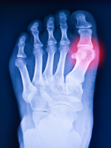
Figure 1: Foot X-ray with Martel’s sign
Case Management
The patient was treated with both drugs, like xanthine oxidase inhibitors, such as allopurinol, as a first-line drug or uricosuric agent, such as probenecid, as a second-line drug. The patient was consulted on maintaining weight within a normal BMI. He did not adopt a crash diet or protein-rich diets such as the Atkins diet. He should integrate reduced-fat foods and vegetarian protein sources more into the diet. He should avoid protein-rich foods like meat, offal, crustaceans, and yeast. The patient must be recommended to increase intake of fluids to more than 2 Liters per day.
Reduced alcohol consumption is recommended, especially for beer and spirits. He should be encouraged to refrain from consuming alcohol for at least three days per week. He was highly recommended for overcoming chronic alcoholism by following any one of the methods, such as psychological counseling, detoxification programme, medications (disulfiram, Naltrexone, Acamprosate), Spiritual practice etc. Beverages with added sugar should be avoided. Consuming soft drinks and drinks containing fructose increased the risk of a gout flare. Because uric acid excretion by the kidneys is inhibited by fructose.
Discussion
In the US, gout affects approximately 9.2 million people. Most diagnoses occur in males and older adults [5]. The number of gout cases is still rising worldwide. In 2017, there were 92 new cases of illness for every 100,000 individuals. This represents an approximate 5.5% rise from 1990 [6]. People of different ethnicities are not equally affected by gout. The populations of Asian Americans and Black Americans have the highest rates, as per the 2021 report [7]. According to research, gout patients who don’t trust their doctors’ advice tend to take their meds less regularly [8]. It’s crucial to educate patients about this and other facets of gout so they know that, despite the possibility of a short-term flare-up, they are working toward a common long-term objective of never experiencing another gout attack [9]. A characteristic of gout is chronicity. It is caused by persistent inflammation that follows gout attacks that recur. Chronic gout can cause cartilage damage, tophi formation, bony erosions, and persistent synovitis. This can be explained by various processes. When urate crystals are present in synovium, chondrocytes are stimulated to produce matrix metalloproteases, nitric oxide, and inflammatory cytokines, which damages cartilage [10]. “Martel sign,” “G-sign,” or “rat-bite” erosion are terms used to describe the radiographic (X-ray) classic finding of gout: a punched-out lytic lesion with a sclerotic margin and an overhanging edge [11]. The characteristic overhanging and displaced bony margin of this radiographic finding is thought to result from erosion of the bone caused by an enlarging tophus from the inside, accompanied by periosteal bony apposition from the outside [12].
As per an initial study investigating the effectiveness of a nurse-led education program it underscores the crucial role of patient education in effectively managing gout. Despite the time-intensive and repetitive nature of patient education, it remains a vital tool for the successful long-term management of gout [11]. The European League Against Rheumatism (EULAR) and the American College of Rheumatology (ACR) have endorsed certain lifestyle recommendations for individuals dealing with gout. These include weight loss for obese patients, avoiding beer (including non-alcoholic varieties), spirits, and sugary sodas, moderating the consumption of meat and seafood, opting for increased intake of skim milk products, and enhancing physical activity. These recommendations stem from epidemiological evidence illustrating the influence of these lifestyle factors on gout risk [1]. It is important to try to avoid using medications that raise uricemia. This mostly applies to medications used to treat hypertension. The average increase in uricemia caused by thiazide and loop diuretics is 0.65 and 0.96 mg/dL, respectively. Increased uricemia and gout risk have also been linked to beta-blockers, non-losartan Angiotensin II receptor blockers (ARBs), and Angiotensin-converting enzyme (ACE) inhibitors. Losartan and calcium channel inhibitors ought to be given preference [13]. Probenecid and allopurinol have long been the mainstays of care for people with chronic gouty arthritis. An appropriate long-term urate-lowering therapy is allopurinol, a xanthine oxidase inhibitor that works by preventing the production of uric acid through a reduction in purine catabolism. Diarrhea, nausea, and elevated levels of ALT, AST, and alkaline phosphatase are common adverse events (AEs) linked to allopurinol. Probenecid is a uricosuric medication that prevents tubular reabsorption to enhance uric acid excretion. Despite being thought to be less effective than allopurinol, it is mostly used as a substitute [14].
Conclusion
This case illustrates that Martel’s sign in chronic gout patients is associated with lytic lesions with overhanging bony edges and sclerotic margins. This clinical condition is well managed by patient education, diet and lifestyle changes, cessation of hyperuricemic drugs, and use of effective Urate-lowering drugs (ULDs).
References
- Ragab G, Elshahaly M, Bardin T. Gout: an old disease in new perspective—a review. J Adv Res. 2017;8(5):495-511. doi:10.1016/j.jare.2017.04.008 PubMed | Crossref | Google Scholar
- Kuo CF, Grainge MJ, Zhang W, Doherty M. Global epidemiology of gout: prevalence, incidence, and risk factors. Nat Rev Rheumatol. 2015;11(11):649-662. doi:10.1038/nrrheum.2015.91 PubMed | Crossref | Google Scholar
- Busso N, So A. Mechanisms of inflammation in gout. Arthritis Res Ther. 2010;12(2):206. doi:10.1186/ar2952 PubMed | Crossref | Google Scholar
- Kumar RR, Jha S, Jain S, Dhir V. Martel sign: a characteristic sign of gouty arthritis. J Clin Rheumatol. 2020;26(7):e217-e218. doi:10.1097/RHU.0000000000001086 PubMed | Crossref | Google Scholar
- Yip K, Berman J. What is gout? JAMA. 2021;326(24):2541. doi:10.1001/jama.2021.19770 PubMed | Crossref | Google Scholar
- Safiri S, Kolahi AA, Cross M, et al. Prevalence, incidence, and years lived with disability due to gout and its attributable risk factors for 195 countries and territories 1990-2017: a systematic analysis of the Global Burden of Disease Study 2017. Arthritis Rheumatol. 2020;72(11):1916-1927. doi:10.1002/art.41404 PubMed | Crossref | Google Scholar
- Butler F, Alghubayshi A, Roman Y. The epidemiology and genetics of hyperuricemia and gout across major racial groups: a literature review and population genetics secondary database analysis. J Pers Med. 2021;11(3):231. doi:10.3390/jpm11030231 PubMed | Crossref | Google Scholar
- Robinson PC, Schumacher HR Jr. A qualitative and quantitative analysis of the characteristics of gout patient education resources. Clin Rheumatol. 2013;32(6):771-778. doi:10.1007/s10067-013-2168-8 PubMed | Crossref | Google Scholar
- Johnston ME, Treharne GJ, Chapman PT, Stamp LK. Patient information about gout: an international review of existing educational resources. J Rheumatol. 2015;42(6):975-978. doi:10.3899/jrheum.141442 PubMed | Crossref | Google Scholar
- Grassi W, De Angelis R. Clinical features of gout. Reumatismo. 2012;63(4):238-245. doi:10.4081/reumatismo.2011.238 PubMed | Crossref | Google Scholar
- De Avila Fernandes E, Bergamaschi SB, Rodrigues TC, et al. Relevant aspects of imaging in the diagnosis and management of gout. Rev Bras Reumatol Engl Ed. 2017;57(1):64-72. doi:10.1016/j.rbre.2016.05.001 PubMed | Crossref | Google Scholar
- Mahajan S, Raina S, Sood V. Tophaceous gout and Martel’s sign: manifestations of advanced gout. APIK J Intern Med. 2022;10(4):278-279. doi:10.4103/ajim.ajim_107_21 Crossref | Google Scholar
Acknowledgments
Not applicable
Funding
Not applicable
Author Information
Corresponding Author:
Raziya Begum Sheikh
Independent Researcher, Department of Content
medtigo India Pvt Ltd, Pune, India
Email: raziya.pharma@gmail.com
Co-Author:
Akshay Hagawane
Independent Researcher, Department of Content
medtigo India Pvt Ltd, Pune, India
Authors Contributions
All patient-related data were collected by Dr. Akshay Hagawane. Raziya Begum Sheikh contributed to the writing of the manuscript, including the original draft preparation and subsequent review and editing to refine the final version.
Informed Consent
Not applicable
Conflict of Interest Statement
This case report is based on a clinical case published in the “Cases” section of medtigo.com. The authors declare no conflicts of interest related to this publication.
Guarantor
Not applicable
DOI
Cite this Article
Raziya Begum S, Akshay H. Martel’s Sign in Chronic Gout. medtigo J Med. 2023;1(2):e3062121. doi:10.63096/medtigo3062121 Crossref


