


 Author Affiliations
Abstract
Glomus tumors (GT) are a rare benign disease that originates from the neuromyo-arterial glomus body, characterized by a triad of aching pain, extreme tenderness, and temperature sensitivity, with the hand being its favoured location. We report a rarely presented GT on atypical site in a 65-year-old female patient who came to the orthopaedic outpatient department with swelling at the medial aspect of the left thigh for three years. It has been increased in size for two years, and it was painless. On examination, it was about 7×9 cm in size, found 6 inches proximally to the medial condyle of the tibia, non-tender, non-fluctuant, erythematous in colour with a white patch over it. Fine needle biopsy reported as GT. Magnetic resonance imaging (MRI) showed the site, size, and expansion to the surrounding structure, such as encasement of the femoral vessels by the tumors. The mass was surgically excised. Biopsy report confirmed encapsulated tumors. A 2-year follow-up, every six months, was carried out for any recurrence, and she was discharged from the clinic at her last follow-up.
Keywords
Glomus tumors, Extradigital, Painless mass, Vascular encasement, Knee, Glomangioma.
Introduction
GT are mesenchymal neoplasms of the glomus body, which regulates blood flow to the skin by changing smooth muscle cells which has the property of contractility.[1] Its anatomy includes the efferent arteriole, anastomotic vessel, primary collecting vein, intraglomerular retinaculum, and a capsule.[2] The pathogenesis of GT remains elusive. However, cumulative evidence suggests several key associations and theoretical underpinnings. Specifically, a robust correlation between GT and neurofibromatosis Type 1 (NF1) has been conclusively demonstrated.[3] This study presents the first comprehensive cytogenetic analysis of GT. Our findings reveal extreme polyploidy in one tumor (NF1-G12) and diploidy in four others. Notably, we identify mitotic recombination of chromosome arm 17q as a novel mechanism underlying biallelic NF1 inactivation in NF1-associated GT.[4]
GT accounting for 2% of soft tissue neoplasms, poses a diagnostic challenge due to its rarity and heterogeneous presentation. The World Health Organization (WHO) classifies these tumors into benign, intermediate glomangiomatosis, and malignant categories. Notably, glomangiomatosis comprises only 5% of cases, with fewer than 50 reported instances of cutaneous malignant GTs worldwide. Further research is warranted to elucidate the pathogenesis and optimize treatment strategies for these enigmatic tumors.[5] Generally, these tumors are most commonly found in females in the hand, while men often experience these cancers in other parts of the body.[6] Mason was the first person to provide the definitive clinical presentation of these tumors, which includes a triad of pain, pinpoint tenderness, and cold sensitivity.[7]
Accurate and immediate diagnosis of these tumors is the preferred way because once it’s diagnosed, then surgical excision of the lesion can give complete symptom resolution in most patients.[8] Several imaging investigations have been proposed to help in the early and rapid identification of these tumors, such as ultrasonography and magnetic resonance imaging (MRI). Ultrasound is the first-line test of choice as it is readily available and low-cost.[9] The histological features of GT depend upon the ratio of vascular to glomus cells, the amount of stroma, and the differentiation of these cells.[10] They are generally regarded as benign, but they can have a malignant and atypical presentation, which features large diameters, multicentricity, and deep locations.[11]
Case Presentation
A 65-year-old, normoglycemic and hypertensive, and hepatitis C virus (HCV) positive female patient presented to the orthopaedic outpatient clinic with a chief complaint of swelling at the medial aspect of the left thigh for the past three years. The patient witnessed that it had increased in size over the past two years and was painless at times. She, however, experienced occasional pain during the cold season. On local examination, it was about 7×9 cm in size, found 6 inches proximally to the medial condyle of the tibia, non-tender to touch, non-fluctuant, erythematous in colour with a white patch over it. Earlier fine needle biopsy, which was done one month back, confirmed the diagnosis of GT, which showed tissue composed of branching vascular channels separated by stroma having glomus cells arranged in nests and aggregates around the blood vessels, with areas of haemorrhages and normal skeletal muscle at the periphery, and there is no evidence of malignancy. All routine lab investigations, such as full blood count (FBC), serum electrolytes, renal function test (RFTs), liver function test (LFTs), and blood sugar level (BSL), were in normal range. MRI clearly proved the site, siz,e and encasement of the femoral vessels by the tumor. The mass was surgically excised in collaboration with the vascular surgeon on board, and the operative findings were an 8x10x7 cm irregular tumor, and the specimen was sent for histopathological test. The report showed that the encapsulated tumor consists of round and oval monotonous cells with pale cytoplasm and a punched-out, rounded nucleus. A 24-month follow-up was kept with the patient for recurrence and malignant potential of the lesion, and fortunately, it did not recur. As a result, the patient was discharged from the clinic.
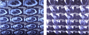
Figure 1: MRI of the lesion
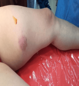
Figure 2: Gross post-operative appearance of lesion
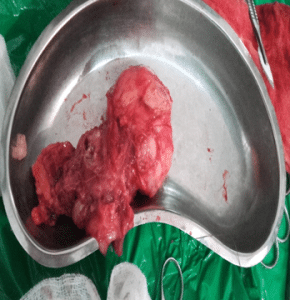
Figure 3: Gross post-operative appearance of lesion
| Lab test | Normal value | Patient’s value |
| Hemoglobin (Hb or Hgb) | 12-16 g/dl | 13 g/dl |
| White blood cells (WBC) | 4-10 X 109/L | 8 X 10⁹/L |
| Creatinine | 0.8-1.3 mg/dL | 0.9 mg/dL |
| Urea | 1.2-3 mmol/L | 1.3 mmol/L |
| Sodium | 136-145 mEq/L | 140 mEq/L |
| Potassium | 3.5-5.0 mEq/L | 3.9 mEq/L |
| Total bilirubin | 0.3-1.0 mg/dL | 0.8 mg/dL |
| Alanine transaminase (ALT) | 10-40 U/L | 30 U/L |
| Aspartate transaminase
(AST) |
10-40 U/L | 35 U/L |
Table 1: Result of Lab tests
Discussion
GT are hamartomas that are most commonly found in the hands in the subungual region (75% to 90%). The aetiology of these tumors is largely unknown, and many hypotheses have been put forward to understand their aetiopathogenesis and the cause of the pain associated with these tumors.[12] It’s a neoplasm found in the skin or soft tissue and is characterized by paroxysmal pain. The normal glomus was described by Hoyer in 1877, and it’s primarily involved in temperature regulation.[4] The clinical trial of paroxysmal pain, pinpoint tenderness, and cold sensitivity is the classic presentation of GT.[13] It has been reported in literature that it can be multifocal (2% to 3 %) and malignant (less than 1 %).[5] The feature of malignant tumors arising in typical GT were found in 60% of the 52 samples studied by Lancerotto L et al. According to their observation, those patients whose tumors met the diagnostic criteria for malignancy were at paramount risk of metastasis and death from the disease. Cases have been reported showing recurrence of the tumors, disease repetition at other parts of the body, and even death as
well.[8]
Extradigital GT are rare entities and thus posing a diagnostic challenge. It has been shown that less than 20% are correctly identified preoperatively. Subsequently, the diagnosis is often delayed. It has been reported that, on average, it took 6.5 years to diagnose GT from the start of the symptoms.[14] Imaging studies to early and accurately diagnose GT are ultrasonography and MRI. MRI is the most sensitive diagnostic modality. Sonographic features of the GT include a hypoechoic, rounded, and well-demarcated mass. MRI will typically show a high signal intensity on T2-weighted images and a low signal intensity on T1-weighted images, which is enhanced with gadolinium.[6] Complete surgical excision is the mainstay of the treatment; however, the presentation of such tumors on a variety of anatomic locations has its own management challenge. If any malignant features are present, then wide surgical resection is indicated with a closed follow-up for recurrence and metastasis. Alternative treatment modalities include sclerotherapy and electron beam irradiation.[15]
Conclusion
There is a general belief that GT affecting the lower extremities is rare. Delayed diagnosis can lead to significant morbidity in terms of pain and discomfort, as seen in our case. Keeping in mind the rarity of this condition, physician should keep a high index of suspicion about GT on extradigital locations such as thighs to diagnose early and to avoid limiting their malignant potential. We reported an extradigital GT arising in the subcutaneous tissue of the thigh. Even though GT is a rare incident and even rarer to find in extradigital locations like the thigh, a review of the literature suggests that these lesions may be more common than they are thought to be, so it should always be kept in mind while attending to patients with such complaints. Surgical resection is the treatment of choice for GT.
References
- Kumar S, Tiwary SK, More R, Kumar P, Khanna AK. Digital glomus tumor: an experience of 57 cases over 20 years. J Fam Med Prim Care. 2020;9(7):3514-3517. doi:10.4103/jfmpc.jfmpc_446_20 PubMed | Crossref | Google Scholar
- Santoshi JA, Kori VK, Khurana U. Glomus tumor of the fingertips: a frequently missed diagnosis. J Fam Med Prim Care. 2019;8(3):904-908. doi:10.4103/jfmpc.jfmpc_88_19 PubMed | Crossref | Google Scholar
- Tang CYK, Tipoe T, Fung B. Where is the lesion? Glomus tumors of the hand. Arch Plast Surg. 2013;40(5):492-495. doi:10.5999/aps.2013.40.5.492 PubMed | Crossref | Google Scholar
- Gombos Z, Zhang PJ. Glomus tumor. Arch Pathol Lab Med. 2008;132(9):1448-1452. doi:10.5858/2008-132-1448-GT PubMed | Crossref | Google Scholar
- Trehan SK, Soukup DS, Mintz DN, Perino G, Ellis SJ. Glomus tumors in the foot: case series. Foot Ankle Spec. 2015;8(6):460-465. doi:10.1177/1938640015583514 PubMed | Crossref | Google Scholar
- Sbai MA, Benzarti S, Gharbi W, Maalla R. A rare case of glomus tumor of the thigh with literature review. J Orthop Case Rep. 2018;8(5):22-24. doi:10.13107/jocr.2250-0685.1192 PubMed | Crossref | Google Scholar
- Liapi-Avgeri G, Karabela-Bouropoulou V, Agnanti N. Glomus tumor: A histological, histochemical and immunohistochemical study of the various types. Pathol Res Pract. 1994;190(1):2-10. doi:10.1016/S0344-0338(11)80490-5 PubMed | Google Scholar
- Lancerotto L, Salmaso R, Sartore L, Bassetto F. Malignant glomus tumor of the leg developed in the context of a superficial typical glomus tumor. Int J Surg Pathol. 2012;20(4):420-424. doi:10.1177/1066896911432454 PubMed | Crossref | Google Scholar
- Morey VM, Garg B, Kotwal PP. Glomus tumors of the hand: review of literature. J Clin Orthop Trauma. 2016;7(4):286-291. doi:10.1016/j.jcot.2016.04.006 PubMed | Crossref | Google Scholar
- Mravic M, LaChaud G, Nguyen A, Scott MA, Dry SM, James AW. Clinical and histopathological diagnosis of glomus tumor: an institutional experience of 138 cases. Int J Surg Pathol. 2015;23(3):181-188. doi:10.1177/1066896914567330 PubMed | Crossref | Google Scholar
- Beksaç K, Doğan L, Bozdogan N, Dilek G, Akgul GGA, Ozaslan C. Extradigital glomus tumor of thigh. Case Rep Surg. 2015;2015:638283. doi:10.1155/2015/638283 PubMed | Crossref | Google Scholar
- Luis LR Jr, Kaoru GR, Shemuel PB, Mills JL Sr. Lower extremity glomus tumors: comprehensive review for surgeons. Vascular. 2008;16(6):326-332. doi:10.2310/6670.2008.00064 PubMed | Crossref | Google Scholar
- Nthumba PM, Oundoh LN. Glomus tumors: a systematic review of the Sub-Saharan Africa experience. Plast Reconstr Surg Glob Open. 2024;12(2):e5564. doi:10.1097/GOX.0000000000005564 PubMed | Crossref | Google Scholar
- Stewart DR, Pemov A, Van Loo P, et al. Mitotic recombination of chromosome arm 17q as a cause of loss of heterozygosity of NF1 in neurofibromatosis type 1-associated glomus tumors. Genes Chromosomes Cancer. 2012;51(5):429-437. doi:10.1002/gcc.21928 PubMed | Crossref | Google Scholar
- Cohen PR. Glomus extradigital tumor: a case report of an extradigital glomus tumor on the wrist and comprehensive review of glomus tumors. Cureus. 2023;15(5):e38737. doi:10.7759/cureus.3873 PubMed | Crossref | Google Scholar
Acknowledgments
We sincerely thank Professor Mohammad Ayaz Khan (Consultant orthopaedic surgeon & head of the department) for his expert guidance and mentorship, elevating this research to excellence.
Funding
None
Author Information
Corresponding Author:
Sohail Rehman
Department of Orthopaedic
Registrar, Global Medics Ireland
Email: sann090909@gmail.com
Co-Authors:
Junaid Zeb
Department of Orthopaedic
NHS Trust UK
Muhammad Ayaz Khan
Department of Orthopaedic
Khyber Teaching Hospital Peshawar, Pakistan
Zia Ullah Jan
Department of Orthopaedic
Khyber Teaching Hospital Peshawar, Pakistan
Authors Contributions
All authors contributed to the conceptualization, investigation, and data curation by acquiring and critically reviewing the selected articles. They were collectively involved in the writing – original draft preparation, and writing – review & editing to refine the manuscript. Additionally, all authors participated in the supervision of the work, ensuring accuracy and completeness. The final manuscript was approved by all named authors for submission to the journal.
Conflict of Interest Statement
The author declares no conflict of interest.
Guarantor
None
DOI
Cite this Article
Sohail R, Junaid Z, Khan MA, Zia UJ. Extradigital Painless Mass of the Thigh: A Rare Presentation of Glomus Tumors. medtigo J Med. 2024;2(4):e30622439. doi:10. 63096/medtigo30622439 Crossref


