


 Author Affiliations
Abstract
Snakebite envenomation has been declared a neglected tropical disease by the World Health Organization (WHO). The only effective treatment of snakebite envenomation is intravenous administration of polyvalent, bivalent, and monovalent snake antivenom. In this study, the comparative in vitro characterization of both Indian (F(ab)₂) and Pakistani (whole immunoglobulin G (IgG)) was carried out. Physiochemical characterization of both antivenom formulations demonstrated a whitish appearance, less than 3% residual moisture, a dry weight of 10 mg per vial, and pH values of 6.89 ± 0.3 for the Pakistani antivenom and 6.96 ± 0.2 for the Indian antivenom. Sterility tests confirmed no bacterial or fungal growth. The protein gel electrophoresis experiment under reducing conditions revealed protein bands between 20 and 35 kDa, corresponding to immunoglobulin light and heavy chains. The Indian antivenom, which consists of F(ab)2 fragments, displayed an additional band at approximately 60 kDa, suggesting the presence of protein aggregates. Contrarily, the Pakistan antivenom, composed of whole IgG immunoglobulins, had an intense band around 20 kDa, indicating the possible presence of co-purified non-specific proteins. F(ab)₂ fragments from the Indian product and complete IgG (~150 kDa) from the Pakistani product were validated by a Matrix-Assisted Laser Desorption/Ionization Time-of-Flight (MALDI-TOF) mass spectrometry study. In both standard pharmacological products, minor peaks linked to tiny serum proteins can be observed. The results demonstrated that both antivenoms are sterile, but the protein aggregates and co-purified proteins increase the risk of adverse immunological reactions. These findings underscore the need for robust quality control and effective purification procedures for antivenoms produced in Pakistan.
Keywords
Snakebite envenomation, Antivenom, Polyvalent antivenom, Protein characterization, Quality control, Protein aggregates.
Introduction
Snakebite envenoming is a relatively unknown tropical disease caused by venomous snakes injecting their specialized, deadly secretions into humans, usually by accident. It has a significant global impact, affecting millions of individuals annually and leading to a substantial number of deaths.[1] Pakistan, located in the northwestern region of South Asia, likewise suffers from high rates of snakebite incidents, with not fewer than 40,000 bites resulting in 1000-8200 deaths every year.[2] The consequences of envenoming vary depending on the snake species involved. Pakistan is home to two families of snakes that include highly venomous species: Elapidae, including kraits, sea snakes, and cobras; and Viperidae, which includes Russell’s vipers and saw-scaled vipers.[3] Antivenom is the only effective remedy for the extensive consequences of snakebite poisoning. It is made by concentrating immunoglobulins from animals that have been hyper-immunized with venom over extended periods of time, such as horses, sheep, and camels. The bulk of Indian polyvalent antivenoms (PAVs) are composed of ammonium sulfate- or caprylic acid precipitated, F(ab)2, which is pepsin-digested fragments of IgG. On the other hand, Pakistan manufactures its own PAV in a single facility.[4] While the purity and physicochemical characterization of several antivenoms obtained from around the world have been assessed, the assessment of the quality and safety of antivenoms and detailed physicochemical characterization from India is uncertain.[5] Each year, the National Institute of Health (NIH) in Islamabad, Pakistan, manufactures about 30,000 polyvalent antivenom vials. However, there isn’t enough antivenom on hand to treat every case of snakebite in the nation.[6] So, snake antivenom sera are currently bought from India to supply the demand for antivenoms. Based on physicochemical analysis, antivenom of snake preparations is constantly noted with purity values exceeding 90%; however, the active material, i.e., the immunoglobulins specific to snake venom, is not assessed by these methods and so might have a purity less than 90%. Most anti-venom producers’ attempts to improve product purity are focused on refining the process of plasma fractionation to produce formulations devoid of non-immunoglobulin protein contaminants. The content of albumin in the final preparations should preferably not surpass 1% of total protein.[7] To improve the physicochemical purity of antivenom, meticulous purification processes are required. All antivenom production recommendations emphasize approaches such as immunoglobulin fractionation and albumin elimination.[8] In this study, we show the possibility and usefulness of using simplified purification and processing methods to improve existing antivenom drugs immediately.
Methodology
Antivenom sample: This research was carried out at the Sera Lab, Biological Production Department, National Institute of Health, Islamabad, Pakistan. Indian snake antivenom was purchased from the Rawalpindi market and locally raised antivenom from the Sera Lab, Biological Production Department, NIH, Islamabad.
Physiochemical characterization of PAV: The physicochemical features of both Pakistani and Indian antivenoms (i.e., color, texture, and uniformity of the product) were evaluated manually by following the WHO guidelines. Vials of both Indian and Pakistani antivenom were examined to check their quality, appearance of lyophilized powder, and presence of moisture. The dry weights of both vials were measured. To check their solubility, the mixtures of each vial were dissolved in 10 ml of de-ionized water. Then the pH of each vial was checked by a potentiometer. The moisture content was determined by the loss on drying method by heating the antivenoms for about 3 hours at 105°C in an oven.[9]
Determination of sterility of snake antivenom: Each snake antivenom underwent a sterility test in accordance with the WHO recommendations. 200 ml of trypticase soy broth and thioglycolate culture media were incubated at 37°C with a milligram solution of PAV to test for bacterial and fungal growth, along with the appropriate control. Blood agar medium for bacterial culture and salted dextrose agar for fungal culture were placed on plates at intervals of 24 hours and 48 hours, respectively. After the plates solidified, 500 ml of culture were removed aseptically and added to bacterial and fungal culture plates. These plates were then incubated at 37°C to measure the bacterial and fungal growth and to look for colonies.
Sodium Dodecyl Sulfate-Polyacrylamide Gel Electrophoresis (SDS-PAGE) analysis of both Indian and Pakistani polyvalent snake antivenom: The electrophoresis was done at room temperature on a mini gel system. 20 µl of both snake antivenoms with a molecular weight range (Broad-Range (10-260 kDa)) were added to 10% and 12% SDS PAGE gels and fractionated under 90 volts for two hours, and resultant proteins were visualized by staining with Commassie Blue R-250. Molecular weights of proteins were determined by analyzing bands and comparing them with bands of a protein marker.
MALDI-TOF analysis of proteins
Requirements for MALDI-TOF: ATCC control 8739 (E. coli) and Pickme Nibs
Sample preparation: Protein samples were purified to a high degree to remove any contaminants or other unwanted substances. Centrifugation of both samples by placing samples in centrifuge tubes in equal amounts. The temperature was adjusted to 37°C by adjusting the rotation per minute (rpm) range to 6000 for 12 minutes. So that the protein could be obtained at a suitable concentration.
Pallet formation through centrifugation: 600 ml of acetonitrile (high-performance liquid chromatography (HPLC) grade) was mixed with 400 ml of serum proteins and centrifuged to obtain protein in the form of a pellet. Supernatant was discarded, and the pellet collected was referred to MALDI-TOF analysis.
Mixing of protein sample with a suitable matrix solution: The protein sample was mixed with a matrix solution known as alpha-cyano-4-hydroxycinnamic acid. This matrix solution helps in the ionization of protein molecules.
MS slide preparation: A Biomerieux Vitek MS slide marked as A-L, a total of 48 rows with three controls, was taken.
Sample spotting: By using Vitek Pickme Nibs, a single isolated colony from an ATCC culture plate was picked up and smeared on the selected circle of control. Using another control, Pickme Nibs 0.1 ml was spread on the MALDI target plate. Here, the dried droplet method was applied. At the end, drying of the MALDI plate was performed.
Insertion of the target plate into the MALDI-TOF mass spectrometer: Targeted MALDI-TOFF plate was put in the mass spectrometer analyzer matrix. Molecules absorbed energy, and ionization of protein molecules occurred, which are present in the sample. Ions that reach the detector (time of flight) depend on their mass to charge (m/z) ratio. The sample contained immunoglobulins and other serum proteins. It took a little bit longer for the identification of large molecular-sized protein molecules.
Mass spectrum identification by dedicated software. The spectrum was obtained, and the resultant PMF was extracted from the MALDI spectrum and searched against different protein databases. We use the Protein Mascot software to demonstrate the search.
Brief description of Protein Mascot: ‘Swiss-Prot database’ From the search list, search e.g., Swiss-Prot. Taxonomy information was put as Equus ferus caballus, which is the origin of the protein sample. Carbamidomethyl (C) was selected as a modification. Oxidation (M) was selected as a variable modification. Peptide tolerance was set to 100-300 ppm. MH+ was selected for mass value. Average was selected for data acquired in linear mode. The spectra file was pasted directly into the Query field. “Auto” we selected from “Report top” hits. The search was started.
Results
Physiochemical assessment of antivenom: Both antivenom drugs appear whitish within the acceptable dry weight range (10 mg per vial). Upon reconstitution, both formulations showed no cloudy appearance, demonstrating good solubility. By fully complying with WHO standards, residual moisture content was less than 3% for both products. The measured pH values were 6.89 ± 0.3 for the Pakistani antivenom and 6.96 ± 0.2 for the Indian antivenom, both falling within the acceptable near-neutral pH range (Table 1).
| No. | Parameter | Locally raised (Pakistani) | Commercially available (Indian) | WHO standards |
| 1. | Appearance | Whitish in color | Whitish in color | Compliance with the description of the marketing dossier |
| 2. | Dry weight | 10 mg | 10 mg | – |
| 3. | Solubility | No cloudiness | No cloudiness | The solution should not be cloudy |
| 4. | Residual moisture content | Less than 3 | Less than 3 | Less than 3 |
| 5. | Sterility Test | No bacterial or fungal growth | No bacterial or fungal growth | No microbial growth |
| 6. | pH | 6.89 ± 0.3 | 6.96 ± 0.2 | Specifications of regulatory agencies and pharmacopeias (generally neutral pH) |
Sterility test: No bacterial or fungal growth was detected for either antivenom on blood agar (for bacterial testing) or Sabouraud dextrose agar (for fungal testing), confirming that both products were sterile.
SDS-PAGE profiling of snake antivenom: Under reducing conditions, snake antivenoms primarily contain proteins above 100 kDa. Proteins were separated under reducing conditions that are consistent with a 20 kDa and 30 kDa mass range for the heavy and light chains of F(ab)2.
Following the reducing condition of SDS-PAGE, protein bands were visualized with a molecular mass range of 20-35 kDa. Additional faint and thick protein bands were also observed. Protein separating at 20 kDa appeared to contain a relatively high amount of that protein. That is consistent with the light chain of the immunoglobulin. In fact, a major band was observed in the 20-25 kDa range. Another band appeared at the MW range between 30 and 35 kDa. Indian snake antivenom contained an additional band at 60 kDa, which can be non-therapeutic or any other protein aggregate, which has nothing to do with venom neutralization of different giant venomous snakes. On the other hand, both Indian and Pakistani snake antivenoms contained additional bands at 15 kDa.
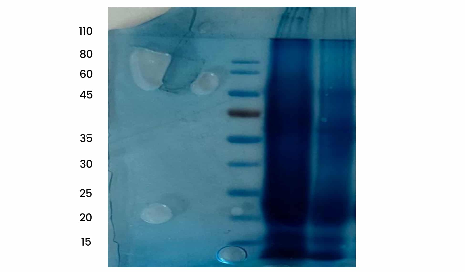
Figure 1: SDS-PAGE Bands
Protein analysis by MALDI-TOF: Through MALDI-TOF analysis of the pellet form of locally manufactured snake antivenom, a spectral peak was obtained that relates to the presence of the major component of that vaccine, known as immunoglobulin IgG, having a molecular mass of 150 kDa. This immunoglobulin contains two light chains and two heavy chains. Through this analysis, only the molecular mass of proteins is identified, and this technique is unable to provide data about the protein sequence. However, through this study, it was revealed that locally available snake antivenom contains whole IgG or intact IgG, and a lot of data or literature is available about the sequence of that immunoglobulin, as each light and heavy chain contains largely similar but not identical amino acid sequences. Each of these chains has 110 amino acid sequences. These light chains are linked with heavy chains through a disulfide bond. Each species contains a varying amount of light chains of immunoglobulin.
Pakistani polyvalent snake antivenom was evaluated in terms of proteomics by MALDI-TOF mass spectrometry spectra obtained after passing the sample through the mass spectrometry analyzer. It was revealed that this antivenom contained a major peak, which obviously confirms the presence of therapeutic antibodies that tend to neutralize toxins and bind their specific antigens. The immunoglobulin showed its peak consistent with molecular weight. Ionization of some proteins and contamination substances occurred. Some other spectral peaks were obtained, which referred to small-sized serum proteins. On the other hand, contamination of the protein sample may have also occurred during sample storage or handling, as there were minor peaks in the spectra of the Pakistani snake antivenom pallet. This suggests sample purification and vigorous laboratory assessment of these therapeutic drugs so that the effectiveness and purity of snake antivenoms are assured at all levels.
Indian polyvalent snake antivenom was evaluated in terms of proteomics by MALDI-TOF mass spectrometry, with spectra obtained after passing the sample through a mass spectrometry analyzer. It was revealed that this antivenom contained a major peak, which obviously indicated the presence of therapeutic antibodies, which tend to neutralize toxins and bind their specific antigens. Derivatives of immunoglobulin showed their peaks consistent with the molecular weight of both light and heavy chains of immunoglobulin. Ionization of some proteins and contaminating substances came to occur. These small-sized proteins have not been effective against venom neutrality. On the other hand, contamination of the protein sample may have also occurred, as their minor peaks in the spectra of Indian snake antivenom pallet. This suggests sample purification and vigorous laboratory assessment of these therapeutic drugs so that effectiveness and purity of snake antivenoms are assured at all levels.
Discussion
This study unveiled the in vitro characterization of both locally and commercially available snake antivenoms. The sterility test confirmed no bacterial/fungal growth or colonies, assuring their safety. Physiochemical tests assured its optimum pH, and a suitable temperature range is assumed for its effective clinical storage. The proteome of both drugs under the reducing conditions verified that Indian polyvalent antivenom contains F(ab)₂ fragments having a molecular range between 20-35 kDa, consistent with immunoglobulin derivatives that contain both heavy and light chains. The presence of a substantial amount of protein aggregate and other bands outside the range of 20-35 kDa is a cause of safety concern, as these non-therapeutic proteins are known as the risk factors of serum reactions and the high cost of treatment. Locally manufactured snake antivenom in Pakistan is a whole IgG form of immunoglobulin, and the Fc portion of that vaccine may cause serum reactions and other complications because, through this electrophoresis and spectrometric analysis, it is suggested that the presence of co-purified proteins must be determined and eliminated before clinical analysis. Moreover, the import of Indian snake antivenom must be limited, and more efforts must be put into in vitro characterization of snake antivenom by employing different and advanced immunochemical and electrophoresis techniques and immunization of model animals with venomous snake species endemic to the local region.
Conclusion
Snakebite envenoming is a major neglected tropical disease declared by the WHO, causing higher rates of deaths and associated disabilities globally. Antivenom is the only effective remedy for the extensive consequences of snakebite envenoming. The efficacy of imported antisera drugs has been challenged, given the venomous snake species endemic to Pakistan. A lack of rigorous in-house laboratory assessment may lead to adverse reactions such as serum sickness. Both the commercially available (Indian) and the locally manufactured (Pakistani) share distinct pharmacological properties while having equal safety and stability features.
References
- Gutiérrez JM, Calvete JJ, Habib AG, Harrison RA, Williams DJ, Warrell DA. Snakebite envenoming. Nat Rev Dis Primers. 2017;3:17063. doi:10.1038/nrdp.2017.63
PubMed | Crossref | Google Scholar - Lim ASS, Tan KY, Quraishi NH, et al. Proteomic Analysis, Immuno-Specificity and Neutralization Efficacy of Pakistani Viper Antivenom (PVAV), a Bivalent Anti-Viperid Antivenom Produced in Pakistan. Toxins (Basel). 2023;15(4):265. doi:10.3390/toxins15040265
PubMed | Crossref | Google Scholar - Hashmi SU, Alvi A, Munir I, et al. Functional venomics of the Big-4 snakes of Pakistan. Toxicon. 2020;179:60-71. doi:10.1016/j.toxicon.2020.03.001
PubMed | Crossref | Google Scholar - Qureshi H, Alam SE, Mustufa MA, et al. Comparative cost and efficacy trial of Pakistani versus Indian anti-snake venom. J Pak Med Assoc. 2013;63(9):1129-1132.
Comparative cost and efficacy trial of Pakistani versus Indian anti-snake venom - Patra A, Herrera M, Gutiérrez JM, Mukherjee AK. The application of laboratory-based analytical tools and techniques for the quality assessment and improvement of commercial antivenoms used in the treatment of snakebite envenomation. Drug Test Anal. 2021;13(8):1471-1489. doi:10.1002/dta.3108
PubMed | Crossref | Google Scholar - Manuwar A, Dreyer B, Böhmert A, et al. Proteomic Investigations of Two Pakistani Naja Snake Venom Species Unravel the Venom Complexity, Posttranslational Modifications, and Presence of Extracellular Vesicles. Toxins (Basel);12(11):669.2020. doi:10.3390/toxins12110669
PubMed | Crossref | Google Scholar - Segura A, Herrera M, Villalta M, Vargas M, Gutiérrez JM, León G. Assessment of snake antivenom purity by comparing physicochemical and immunochemical methods. Biologicals. 2013;41(2):93-97. doi:10.1016/j.biologicals.2012.11.001
PubMed | Crossref | Google Scholar - Tan CH, Liew JL, Chong HP, Tan NH. Protein decomplexation and proteomics: A complementary assessment method of the physicochemical purity of antivenom. Biologicals. 2021;69:22-29. doi:10.1016/j.biologicals.2020.12.004
PubMed | Crossref | Google Scholar - Ahn JY, Kil DY, Kong C, Kim BG. Comparison of Oven-drying Methods for Determination of Moisture Content in Feed Ingredients. Asian-Australas J Anim Sci. 2014;27(11):1615-1622. doi:10.5713/ajas.2014.14305
PubMed | Crossref | Google Scholar
Acknowledgments
The author thanks the staff of the Sera Lab, Biological Production Department, National Institute of Health, Islamabad, Pakistan, for offering technical support and laboratory facilities for this research.
Funding
There is no external/internal funding available.
Author Information
Aswad Khan
Department of Biological Sciences
International Islamic University, Islamabad, Pakistan
Email: [email protected]
Author Contribution
Aswad Khan is the sole contributor of this manuscript. He conceptualized the study, designed the methodology, carried out all laboratory work, analyzed the data, and wrote and revised the manuscript.
Ethical Approval
Not Applicable
Conflict of Interest Statement
There is no conflict of interest.
Guarantor
Aswad Khan is the guarantor of this author and holds full responsibility for the integrity of the data and accuracy of the study.
DOI
Cite this Article
Khan A. Comparative In-Vitro Characterization of Commercially Available and Locally Raised Polyvalent Snake Antivenom. medtigo J Pharmacol. 2026;3(2):e3061323. doi:10.63096/medtigo3061323 Crossref


