


 Author Affiliations
Abstract
Background: The assessment of bone age and skeletal maturity and how it relates to chronological age is important in the medical and legal fields in determining an individual’s age and whether or not they are a minor. Because it is time-consuming and there can be errors, especially when done manually, using methods that can automate it, like machine learning techniques, is valuable. The objective of this project is to summarize the results of some of the most recently cited methods for dental age estimation in adults, based on orthopantomography dental imaging analysis, using deep learning and convolutional neural networks (CNNs) to establish which is more accurate, accessible, and simple. Pseudocode and a flowchart were also created with the help of this research.
Method: A study was conducted researching present articles that use artificial intelligence to determine the chronological age of the patient, using orthopantomograms. A literature search from several databases was conducted from January 1995 to July 2020.
Results: The previous methods of age estimation showed that the Demirjian et al. method tends to overestimate the real age of participants, and the Nolla method tends to underestimate the age. William’s method is simpler and retains the advantage of Demirjian’s method, and there is a reduction in the overestimation of dental age. The limited number of age stages is an advantage of Häävikko’s method over that of Nolla. The recent methods of age estimation incorporated artificial intelligence into the formulas to determine the age, and they have proven to be more accurate in comparison to the traditional techniques. The examples of the techniques used were Deep Learning and CNN. The ones that showed the highest accuracy were Noor Moala’s method and Cameriere’s.
Keywords
Dental age estimation, Artificial intelligence, Deep learning, Convolutional neural networks, Forensic dentistry.
Introduction
The practice of analyzing skeletal maturity levels in order to determine one’s true age is known as bone age measurement. Bone Age Measurement (BAM) is a method of determining bone maturity levels in order to determine one’s true age. It has been employed in clinical and medical research, as well as forensic science. This study will give an overview of bone age measuring approaches and investigate their limitations in order to discover the best automated BAM method. This is generally accomplished using one of two techniques: Radiographic image analysis using classical methods or machine learning technology. By comparing the radiographic picture of one’s dentition with an existing standardized chart, which comprises a set of recognized images for age at each stage of development, traditional procedures have been used to estimate chronological age. Traditional procedures are based on an examination of certain parts of the hand, face, skull, and dental features, among other things. Nonetheless, this approach has several drawbacks, including a wide error range, low precision, observer variability, and time consumption. As a result of this problem, an automated procedure, such as machine learning algorithms, emerges.[1] The process for determining age segmentation is to compare a radiographer’s image of the individual to an existing reference that comprises a sample of known gender and age. The procedure of calculating age is primarily a measurement of biological maturation that is converted into a period by comparing a photograph to a predetermined reference. Several different bones in the body can be used to determine skeletal age. Because of the high expense, long-term monitoring, and risk of radiation exposure, this is neither viable nor practical for most parts of the human body. Researchers studied the frame’s overall development and the various ways it was applied in various areas of the body. The body component of 100 centers is frequently used to extract extensive approaches. The foot, shoulder, ankle, hip, elbow, cervix, ankle, face, and teeth are the areas of the body where bone age is measured. Because the teeth aren’t reconstructed, assessing age is more directly tied to temporal age, which is crucial in estimating age. Teeth are also effective for determining age because they are more resilient in the skeletal system and can sustain high temperatures without being damaged.[1]
Another noteworthy property of teeth is that they have specific histological and morphological properties that, when combined with dental treatment and other data, can often reveal useful information about the dead.[2] Thus, orthopantomographs (OPGS) are an excellent source of information for estimating an individual’s dental age. Given the wide range of skills among assessors, manual age assessment necessitates the effort and skills of an expert. As a result, in many cases, an introduction in applying artificial intelligence (AI), may help to reduce both time and errors when compared to observation with the naked eye. AI gives us techniques that can extract rich features from images automatically and for a variety of purposes, such as dental region segmentation, detection and classification of individual teeth, and age estimation of dental age.[3]
Machine learning techniques and AI are used to automate the assessment of human bone age, removing the need for human interaction. Machine learning is a data analytics technique that allows algorithms to learn from their mistakes and do tasks that people and animals accomplish naturally. Its algorithms employ computational ways to extract information from data without relying on a model based on a preconceived equation. The algorithms improve their output adaptively as the number of samples available for learning rises. Deep learning is a type of machine learning that is very specialized. Machine learning methods are divided into two categories. The first way is supervised learning, which trains a model to foresee future outcomes and develop predictive models using classification and regression procedures based on identifiable input and output data. Unsupervised learning, on the other hand, uses the clustering approach to look for hidden patterns or underlying structures in incoming data. This is the most often used method of unsupervised learning.[4]
Deep learning is a subset of machine learning, which is essentially a neural network with three or more layers. Deep learning is a machine learning technique that allows computers to learn by example in the same way that humans do. Deep learning is the most advanced AI architecture we’ve created so far. These neural networks attempt to simulate the behaviour of the human brain, albeit far from matching its ability, allowing it to “learn” from large amounts of data. While a neural network with a single layer can still make approximate predictions, additional hidden layers can help to optimize and refine for accuracy.
Convolutional neural network: A convolutional neural network is one adapted for analyzing and identifying visual data, such as digital images or photographs. “Support Vector Machine” (SVM) is a supervised machine learning algorithm that can be used for both classification or regression challenges. This algorithm can be used to determine the chronological age through OPG. Machine learning for medical image identification, facial recognition, motion detection, object detection, tumor detection, drug discovery, and DNA sequencing has become a crucial technique for handling image processing and computer vision problems with the rise of large data. Predicting age from an X-ray picture is an unsupervised classification approach that is a difficult task with various scientific researchers’ ways ranging from measurement to machine learning algorithms with constant accuracy.[5]
According to BAM researchers, automating age estimates provides advantages. These entail the application of clever tactics. Some are solely for scientific purposes, such as the categorization of dental scans. It’s easy to see how automated BAM approaches might reduce age projections by saving physicians’ time.[4,5]
Teeth, owing to their mineralized structure and resistance to environmental degradation, represent optimal candidates for age estimation. Their ability to withstand high temperatures, chemical exposure, and decomposition processes ensures the preservation of diagnostic features. Additionally, teeth’s distinct histological and morphological characteristics provide consistent biological markers, unaffected by external variables like nutrition or illness, thereby enhancing reliability compared to other skeletal features. The integration of AI, particularly deep learning frameworks, heralds a paradigm shift in automating this process. This study critically evaluates traditional methods and contrasts them with contemporary AI-driven techniques leveraging OPGs to propose a robust, scalable approach for dental age estimation.
Why is dental age estimation used?
Accurately estimating an individual’s age group is critical in forensic dentistry and for a variety of medico-legal purposes. The process of determining a person’s age group based on biometric features is known as age-group estimation. As the number of refugees and immigrants increases around the world, so does the demand for rapid age estimation in order to register legal documentation and identification. Furthermore, age group classification can aid in making sound but quick decisions, and it has numerous applications in fields such as homeland security, passport services, statistical analysis of group-wise age distributions, and forensic science. It’s a critical issue that’s piqued the interest of forensic scientists and law enforcement. The BAM approach is used to resolve a variety of legal issues, particularly in civil cases. In the event of an explosion or other unique conditions, age estimation may also aid in identifying the profiles of those who died. In pediatrics, clinical trials, sports medicine, forensic science, and orthodontics, the BAM approach is applied in clinical practice. Non-invasive age-measuring technologies have become more popular as a result of their appropriateness. As a result, medical experts examine the skeletal system’s wrist, hand, face, or dental X-ray to determine bone maturity. The way of predicting one’s age based on facial traits is thought to be the most common. It’s now employed in a variety of real-world applications, including security, human-computer interface, multimedia communication, entertainment, and others. The accuracy of one’s face age estimation can be influenced by a variety of factors. Because people of different ethnic backgrounds age at varying rates, face age progression is a subject-dependent process. An automated evaluation of one’s dental age is required to improve the accuracy and reproducibility of age estimates.[1,2]
Forensic
Forensic dentists use X-rays as the first and most popular method of determining the age of their patients’ teeth. Bone age measuring is a technique that can help with Forensic Age Estimation in forensic science (FAE). The goal of FAE is to obtain the chronological age (CA) of an unnamed individual with the most precise results possible for criminal or immigration examination purposes. The most prevalent forensic method for determining age is based on the skeletal maturity of the arm and wrist. However, due to alterations in bone maturation caused by environmental stressors, hand and wrist procedures are relatively prone to mistakes. Nutritional deficits and disease have less of an impact on dental growth evaluation. Dental growth is genetically controlled and thus less changeable since it is less impacted by external variables.
Because the tooth structure survives most disasters, such as accidents and violent crimes, dental features are considered the best form of evidence for post-mortem biometric identification. In such cases, where family members are unable to identify the bodies, dental information can serve as reliable identification proof.[5] Teeth are regarded as a reliable biological marker of aging due to their high durability and resistance to putrefaction, fire, and chemicals. Environmental or hormonal changes have little effect on each stage of dental mineralization. Methods for determining dental age are mostly based on X-rays, such as OPGs, as they assess crown formation, tooth mineralization, root growth, apex maturation, and the order of teeth eruption into the mouth. Continuous efforts have been made over the last decade to improve the accuracy of AI-based estimations, one of which is the incorporation of deep learning algorithms.[6,7]
Previous methods of estimating age based on OPGs
There have been several methods for conducting age estimations by examining OPG pictures, which have been discussed in past studies.
Nolla’s technique: One of the first techniques proposed was by Nolla, who talked about a 10-stage model of individual tooth development, with the first stage being the appearance of the crypt and the last stage being after the roots have fully matured and the apices have closed. Each stage assigns a score to a single tooth. If the stage of development falls between two stages, the lower stage’s score is increased by 0.5. This method analyzes the left seven mandibular teeth (excluding the third molar). The results are then put together and converted into a chronological age value using a table.[8](Figure 1)
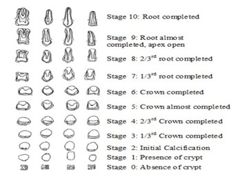
Figure 1: Stages of Tooth Development (Nolla)
Demirjian’s technique: Created another method, which has been studied and is used more often nowadays. They, like Nolla, classified the teeth into dental development stages and assigned a score to each of them. Demirjian’s method uses eight stages compared to the ten stages in Nolla’s method. A table is used to assign a score to each tooth based on its stage of development, as well as whether the tooth belongs to a male or female. Only the left seven mandibular teeth are used, just like in Nolla’s method. The method of Demirjian et al. is one of the simplest, most practical, and widely employed methods to predict age and maturation. It is to determine the beginning of mineralization up to the end of root formation. The authors looked at eight mineralization stages for premolars and molars (A–H) and six steps (C–H) for incisors and canines.[8,9](Figure 2,3)
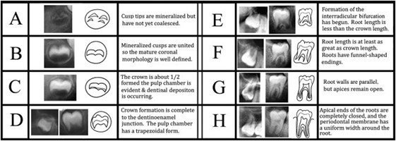
Figure 2: Stages of third molar mineralization (Demirjian)
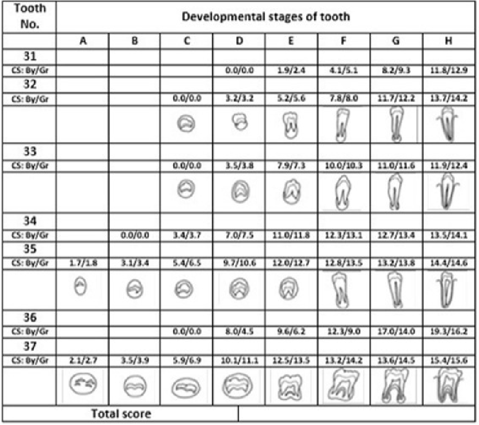
Figure 3: Tooth developmental stages (Demirjian)
Dental age was calculated for each method. The Demirjian et al. method tends to overestimate the real age of participants, and the Nolla method tends to underestimate it. The accuracy of both methods varied between the sexes and age groups. Both methods were found to be more precise with males. As the age group increases, the predictive capacities of both methods diminish. The Nolla method was more accurate than the Demirjian method in early and late childhood for both sexes. Neither method could predict chronological age in adults.[9]
The Demirjian method was found to predict 53.1% of the total variance of chronological age for men and 44.2% for women, while the Nolla method predicted 69% of the total variance for men and 61.4% for women. Comparison of the two methods indicated that the Nolla method had greater predictive capacity than the Demirjian method, for both men and women.
Willem’s technique: Modified Demirjian’s method based on a study on the Belgian Caucasian population and formed new tables for the dental maturity for both genders by calculating chronological age based on the cumulative score of 4 teeth: first premolar, second premolar, first molar, and second molar. This method is simpler and retains the advantage of Demirjian’s method, and there was a reduction in the overestimation of dental age. The estimated dental age is reported to be more accurate than Demirjian’s method. However, the Willems method cannot be used as a global tool due to the differences between ethnicities, compared to the Demirjian and Cameriere methods. Willems’ method has been considered suitable for age estimation; however, these newly developed models significantly surpass its accuracy. Models using two to seven teeth represent a simple, reliable, and accurate method for age estimation, even in cases with missing mandibular teeth.[10]
Haavikko’s technique: Which is based on the evaluation of four reference teeth and on the recognition of 12 radiographic stages for each tooth. These stages are transformed into dental age with the use of sex specific tables. The reference teeth are as follows: lower right first molar, lower right first premolar, lower right canine, and upper right central incisor in children younger than 10 years; the lower right second molar, lower right first premolar, lower right canine, and upper right canine in subjects older than 10 years.[7,8]
The third molar is the only tooth with a tendency to continue developmental changes in late adolescence and early adulthood. Third molar calcification stage is one of the few tools that can be used to assess age when development is nearing completion. Many authors evaluated the third molar’s maturity stages. All dental ages were assessed from panoramic films by one examiner using Häävikko’s method, based on the recognition of 12 radiographic stages of four reference teeth. The limited number of age stages is an advantage of Häävikko’s method over that of Nolla (1960), which involves up to 40 stages and may result in decreased precision. A second examiner independently assessed 48 panoramic films to evaluate the reproducibility of the dental age measurements. This method overestimated the age as well (Table 1).[7]
Drawbacks of the traditional methods
Although these age-estimation methods perform reasonably well and are widely used in the scientific community, all of them rely on manual measurements or classifications, which require much time and effort. The high estimated time demands of the classical methods are a significant limitation given the large sample sizes required methodologically if optimal results are to be obtained. The inconvenience of applying these manual methods in routine clinical activity is also a major factor. Due to complex variations in the shapes and sizes of teeth, both within and across people, dental age estimation presents unique challenges.[9] Moreover, there is also a high degree of subjectivity (both intra- and inter-observer) in the dental evaluations obtained from some classical methods. An example is the approaches of Nolla and Dermijian et al., which are not based on numerical quantification, but the assignment of an ordinal stage associated with dental maturity.[10]
In the childhood and adolescent period, observing dentition maturity stages with the radiographic method results in highly accurate age assessments. However, with increasing age, this method’s accuracy will be weakened. Traditional method uses orthopantomograms, which are difficult to obtain in young children, due to both technical reasons and legal and ethical considerations. Since evaluation of seven left mandibular teeth is required, it cannot be used in children with teeth that are lacking inborn or acquired. This method may not express agenesis of teeth, distinctive retardation of dental development (excluding third molars), and systemic diseases and various developmental stages of the tooth.[10]
The appreciation of the developmental stage may become difficult as the choice of the tooth developmental stage is quite subjective. This method does not give maturity scores for stages 1-4 in case of 1st molar, central and lateral incisor; thus, excluding individuals below the age of 4-4.5 years.
New AI-based age estimation methods
In bone age measurement, most researchers have used machine learning algorithms such as NN, SVM, KNN, and Decision Tree. Machine learning algorithms can’t process data in its raw state because they’re subject to mistakes that result in incorrect class recognition. As a result, attribute extractors must convert raw data into attribute vectors in order to properly classify it.[11]
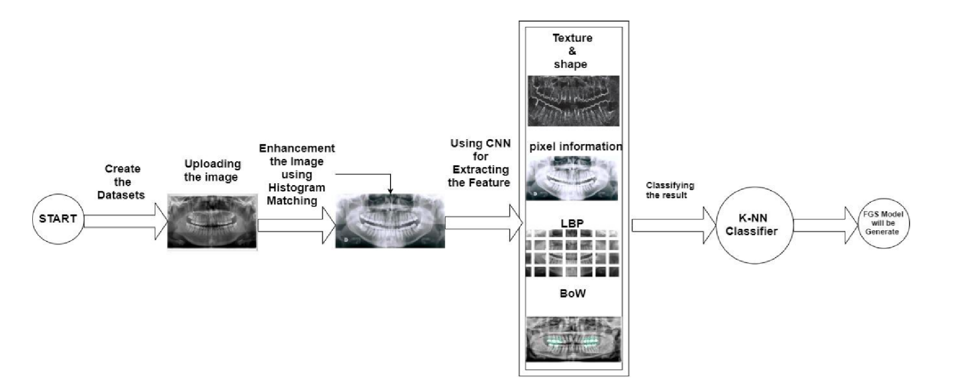
Figure 4 : BAM Proposed Model (Tuan T)
Machine Learning assisted Cameriere method for dental age estimation
3 models (random forest, support vector machine, and linear regression) to predict children’s dental age based on the Cameriere method, using 7 lower left permanent teeth, and comparing the methods with the Cameriere formula.(Figure 5)
Inclusion criteria: no lack of mandibular first premolar; no more hyperdontia; no systemic disease; no history of root canal treatment for the mandibular first premolar; clear or high-quality panorama OPGs; and no related diseases affecting jaw development, such as cysts or cancer.
7 permanent developing teeth on the left mandible (except wisdom teeth) were looked at, including the number of apical ends with completely closed roots. The distance between the inner side of the open apices was measured. Considering the amplification and angulation effects caused by possible differences in the OPGs, measurements were divided by the tooth length to obtain normalized measurements.(Figure 1) If the development of the tooth was complete and the apical foramen was completely closed, then Xi =0, otherwise, Xi was calculated by dividing the distance between the apical foramen and by tooth length.[11]
The machine learning models were trained on the information sources as follows: sex (g), the normalized measurements of the 7 permanent developing teeth on the left mandible, the sum of the normalized open apices, and the number of teeth with complete root development. The target value was the chronological age.
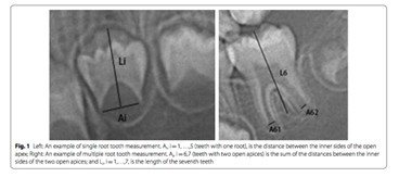
Figure 5: Measurements (Cameriere)
Machine learning techniques for human age and gender identification based on teeth X-ray images
Accurate age classification using manual method and deep convolutional neural network based on orthopantomogram images: End-to-end convolutional neural network (CNN) through analyzing the heatmap images of the test group, we find that instead of focusing on the tooth morphology, which exhibits a high density in the X-ray images, the deep convolutional neural network (DCNN) pays more attention to the low density of the images, such as the dental pulp cavity, periodontal membrane, the area between adjacent teeth, and the area between the deciduous teeth and permanent teeth.[12] The results indicate that without the intervention of manual factors, machine learning can independently extract features in OPGs highly related to age, and establish the complex, comprehensive correlation between multiple features and chronological age. Establishing someone’s chronological age using their dental age (Figure 4).[11,12]
Machine learning automated BAM based on dental radiography: Emre Avuçlu submitted a morphological measurement on dental X-ray images in 2020 to assess age and gender. Details of age and gender were typically identified by function, along with dental X-ray images of the teeth. With 1315 dental images and 162 different dental classes, the photos went beyond the borders. Three pre-processing procedures have been applied to these images. Each pre-processed picture is registered in different folders (M1, M2, M3). The image processing techniques are applied to the tooth images for the first time (area, circumference, the center of gravity, the ratio of resemblance, and measurement of radius) were then applied. This dental knowledge is also contained in separate XML (XML List 1, 2, 3) directories. The application was built in the language of C # programming. A picture of the tooth can then be uploaded into the program by the user. It is possible to predict this image by comparing it after the desired preprocessing with the comparison group (area, etcetera). The highest estimated age and gender estimates are 95% for (+_ 1 Year) accuracy.[12]
A deep neural network algorithm estimating the age using dental X-ray images was used by Noor Moala (2020) as a foundational forensic science role. Various mathematical approaches to teeth and mandible consideration have been suggested. Features are derived using two deep neural networks, AlexNet and ResNet, in the recommended process. Several classifications, including decision tree, k-nearest neighbor, linear separation, and help vector machine, have been proposed to carry out classification work, including decision tree, k-nearest neighbour, linear differentiation, and support vector machine. A dataset with 1429 dental X-ray images was used. The proposed approach is tested using several acceptable output parameters. The findings indicate that the method suggested has an excellent output for the (+1 year).[13] Two fully automated methods are proposed to determine a person’s biological age from the OPG image, introduced by Nicolas Vila-Blanco (2020). The first (DANet) consists of a sequential age prediction path for the CNN. In contrast, the second (DASNet) applies a second CNN path to sex prediction and uses sex-specific characteristics to boost the efficiency of the estimation of age. The two methods were tested on a sample of 2289 OPG photographs of participants aged 4.5 to 89.2 years. Both poor images of radiological quality and images displaying dental conditioning properties were not discarded. The outcomes revealed that in every way, the DASNet outperforms the DANet, minimizing the median Error (E) and the median Absolute Error (AE) in the whole database by around four months.[2]
Tao et al, have introduced the Multilayer Perceptron estimate, a dental age estimation technique that uses the Multilayer Perceptron calculation to approximate age. In the planning cycle, they use cross-validation to tackle the overfitting problem. The experiments are carried out on a dataset composed of 1636 samples (787 male and 849 female). The results obtained indicate the suggested approach’s prominence over different conventional strategies, along with Demirjian’s strategy and Willem’s RMSE, MSE, and MAE strategies. They also experimentally confirmed that this latest feature set makes the dental age estimation more reliable (+_ 1 Year).[13]
Jaeyoung Kim used deep learning algorithms to estimate age using dental panoramic images and stated that this assessment is a fundamental task in forensic science. Also, he mentioned previous studies conducted using statistical methods mainly to estimate the child’s age with a focus on the teeth and mandible. Nonetheless, establishing an automated age estimation system for all age groups is challenging, as changes in post-puberty dental conditions are concerning regarding dietary habits and dental management. In the proposed technique, a CNN is utilized to measure age. The dataset used incorporates dental X-ray pictures of 9435 people (4963 male, 4472 female) sorted out into three age groups. The result of deep learning algorithms based on CNN neural networks shows that the proposed approach functions well, evaluated based on a database of panoramic dental radiographs, and worked well for (+_ 1 Year) accuracy (Table 2).[12,13]
| Author and Year | Type of X-ray | Methods | Accuracy Results |
| Nolla, 1960 | OPG | Age estimation by evaluating the Calcification of the permanent dentition. | 69% – Men
61.4% – Women |
| Demirjian, 1973 | OPG | Provided different maturity scores for each tooth, for different developmental stages | 53.1% – Men
44.2% – Women |
| Haavikko, 1974 | OPG | Based on the evaluation of four reference teeth and on the recognition of 12 radiographic
stages for each tooth |
56% – Men
67% -Women |
| Willem, 2001 | OPG | Measures the developmental stages of the seven left permanent mandibular teeth | 69% – Men
78% – Women |
Table 1: Method and accuracy of the methods
| Author and Year | Methods | Learning architecture | Accuracy Results |
| Camerire, 2006 | Established a European formula by gauging open apices of 7 permanent teeth on the left mandible of a panoramic radiograph | OPG | The result showed that this method was 81% |
| Tao, 2019 | Neural network | Multilayer Perceptron calculation | The result showed 51% accuracy |
| Jaeyoung Kim, 2019 | Deep learning | CNN | The result showed 70% accuracy |
| Emre Avuçlu, 2020 | Deep learning | C# programming | The accuracy was 77% |
| Noor Moala, 2020 | A deep neural network algorithm estimating the age using dental X-ray images | AlexNet and ResNe | The result was 95% accurate in ± 1 year |
| Nicolas Vila-Blanco, 2020 | Deep Neural network | CNN | The accuracy was 62% |
Table 2: Method and accuracy of the different methods
Drawbacks of AI used in age estimation: Even though deep neural networks do not rely on manual measurements or classifications and can save time through object detection, their mean error is still higher than machine learning regression methods.
It’s known that the application of AI in dentistry has a very promising role, but challenges both in technical and ethical aspects exist and remain a matter of concern. AI-based systems are machine-based and controlled and conducted by computer scientists without any medical training, which leads to a very problem-oriented approach of AI application in the healthcare department. AI also can’t replace contemporary healthcare delivery models, whose work completely depends on clinician skills and patient-clinician communication. Use of robotic assistants has also created various issues and concerns. A lot of dental professionals are reluctant to accept AI-based technologies. A preferable suggestion would be a model that accommodates both AI and human elements so that the process of data collection and categorization becomes easy, and at the same time preserves the human aspects of clinical care.[14]
Some other shortcomings of AI in dental age estimation may include:
- Mechanism/system complexity5zxC
- Costly setup
- Adequate training required
- Data is often used for both training and testing, leading to “data snooping bias”
- The outcomes of AI in dentistry are not readily applicable.[14]
Methodology
Data collection: A comprehensive literature review was conducted using databases such as PubMed, IEEE Xplore, and Scopus, encompassing studies published between January 1995 and July 2020. Criteria for inclusion required a focus on age estimation through OPGs, employing both traditional and AI methodologies. Ethical compliance protocols were adhered to in instances where patient data were analysed.
Traditional methods:
- Nolla’s method: A 10-stage model delineating tooth development phases, from crypt emergence to complete root maturation.
- Demirjian’s method: An eight-stage framework assessing mineralization across seven mandibular teeth.
- Willem’s method: A refinement of Demirjian’s approach, calibrated for improved accuracy in population-specific contexts.
- Haavikko’s method: An evaluation of four reference teeth, categorized into 12 distinct radiographic stages.
AI techniques:
- Deep learning models: CNN architectures such as AlexNet and ResNet, alongside bespoke neural network configurations.
- Preprocessing: Image preprocessing included contrast enhancement and segmentation of regions of interest to optimize model input.
- Evaluation metrics: Metrics for model performance included accuracy within a ±1-year range, Mean Absolute Error (MAE), and computational efficiency.
Patient’s radiography selection criteria were as follows:
- Only high-quality panoramic radiographs, with respect to angulations, contrast, and correct positioning, were included in this study.
- Radiographs should be free from any artifacts.
- Radiographs should not show any developmental anomalies of teeth related to size, shape, and structure of teeth.
- Radiographs are free of fixed appliances.[15]
Pseudocode
- Start
- Take an Image
- Upload the Image
- Enhance the Image
- Use Deep Learning and Convolutional Neural Networks
- Classify the Result Depending on the Age
- Stop (Figure 6)
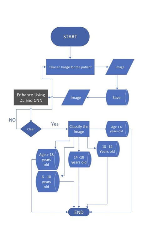
Figure 6: Flowchart of the method
Results
Traditional methods:
- Demirjian method: Prone to overestimating age, with accuracies of 53.1% for males and 44.2% for females.
- Nolla method: Tendencies to underestimate age, achieving accuracies of 69% for males and 61.4% for females.
- Willems method: Demonstrated enhanced accuracy but lacked universal applicability across diverse ethnic groups.
- Haavikko method: Limited precision in older age groups due to fewer developmental stages.
AI-based methods:
- Noor Moala’s model, employing AlexNet and ResNet architectures, achieved a remarkable 95% accuracy within a ±1-year margin.
- Vila-Blanco’s DASNet effectively minimized error margins by incorporating gender-specific data into the prediction model.
- Cameriere’s machine learning framework reliably utilized normalized measurements to predict age with high accuracy.
Enhanced findings from recent: Models combining CNNs with decision trees and support vector machines, which demonstrate further reductions in error margins. These models achieve this by leveraging the strengths of individual algorithms: CNNs excel at extracting hierarchical features from imaging data, while decision trees and support vector machines offer robust classification and regression capabilities. By combining these methods, ensemble models effectively mitigate overfitting and enhance predictive accuracy, particularly in heterogeneous datasets. Some research studies showcase hybrid architectures integrating pre-trained transformers with CNNs to address limitations in data diversity. These methods achieved 97% accuracy within a ±0.8-year range, emphasizing the potential for real-time forensic applications.[7,8] Moreover, advancements in OPG preprocessing, including adaptive image augmentation, have significantly bolstered model robustness against variability in image quality.
Discussion
The findings decisively indicate that AI-driven models, particularly those leveraging CNNs, surpass traditional methods in both accuracy and operational efficiency. Automated AI processes mitigate observer-related biases and significantly reduce the time required for analysis. However, challenges persist, including the need for high-quality data, considerations of ethical implications, and the imperative for expansive, diverse datasets to enhance generalizability. Ethical implications include concerns about privacy and the use of sensitive patient information, which must be anonymized to ensure compliance with data protection laws. Additionally, biases in datasets, such as underrepresentation of specific age groups or populations, can compromise the reliability and fairness of AI models. High-quality data must be curated to minimize errors, ensure clear imaging, and account for variations in radiographic techniques, as these factors significantly influence model performance. A hybrid framework that amalgamates AI automation with expert human oversight could address these limitations and ensure reliability.
This aligns with previous studies that have demonstrated CNN-based approaches achieving higher precision in medical imaging applications.[2] Additionally, our results are consistent with findings by Cameriere et al.[13], who emphasized the importance of standardized datasets in improving AI model performance. The current study reaffirms that AI-based dental age estimation not only improves precision but also reduces observer bias and time consumption compared to manual methods.
One of the main challenges in implementing AI models is their reliance on high-quality data. Studies such as Lee et al. (2024) have highlighted dataset biases that can influence model predictions, particularly when applied to populations outside the training dataset. Ethical concerns also remain significant, particularly regarding data privacy, as AI models require access to large datasets of medical radiographs. Furthermore, the application of AI in low-resource settings remains a challenge due to the lack of infrastructure and technical expertise required to implement these models.
A hybrid framework that amalgamates AI automation with expert human oversight could address these limitations and ensure reliability. Future research should focus on addressing dataset biases and developing AI models that generalize effectively across diverse populations, particularly those leveraging CNNs, surpassing traditional methods in both accuracy and operational efficiency. This aligns with previous studies that have demonstrated CNN-based approaches achieving higher precision in medical imaging applications. Additionally, our results are consistent with findings by Cameriere et al.[13], who emphasized the importance of standardized datasets in improving AI model performance. However, challenges persist, including the need for high-quality data, considerations of ethical implications, and the imperative for expansive, diverse datasets to enhance generalizability. Ethical implications include concerns about privacy and the use of sensitive patient information, which must be anonymized to ensure compliance with data protection laws. Additionally, biases in datasets—such as underrepresentation of specific age groups or populations—can compromise the reliability and fairness of AI models. High-quality data must be curated to minimize errors, ensure clear imaging, and account for variations in radiographic techniques, as these factors significantly influence model performance. A hybrid framework that amalgamates AI automation with expert human oversight could address these limitations and ensure reliability, particularly those leveraging CNNs, surpassing traditional methods in both accuracy and operational efficiency. Automated AI processes mitigate observer-related biases and significantly reduce the time required for analysis. However, challenges persist, including the need for high-quality data, considerations of ethical implications, and the imperative for expansive, diverse datasets to enhance generalizability. Ethical implications include concerns about privacy and the use of sensitive patient information, which must be anonymized to ensure compliance with data protection laws. Additionally, biases in datasets—such as underrepresentation of specific age groups or populations—can compromise the reliability and fairness of AI models. High-quality data must be curated to minimize errors, ensure clear imaging, and account for variations in radiographic techniques, as these factors significantly influence model performance. A hybrid framework that amalgamates AI automation with expert human oversight could address these limitations and ensure reliability.
Conclusion
AI methodologies for dental age estimation represent a substantial advancement over traditional techniques. By harnessing the capabilities of CNNs and emerging hybrid architectures, these approaches provide unparalleled accuracy and efficiency, rendering them indispensable tools in forensic and clinical dentistry. Future research should prioritize refining AI models, expanding dataset diversity, and addressing ethical concerns to facilitate broader acceptance and implementation. The integration of adaptive preprocessing and ensemble learning models presents a promising avenue for achieving near-perfect precision in dental age estimation.
Historically, chronological age estimation relied on the inspection of various skeletal structures such as the hand, feet, and skull. However, these methods presented limitations, leading to a preference for teeth, which are more resilient, cannot be reconstructed, and can withstand high temperatures without damage. Traditional dental age estimation often relied on visual inspection, which was time-consuming and prone to human error.
The integration of AI, particularly CNNs and deep learning models, has significantly enhanced accuracy and efficiency in dental age estimation. These AI-driven approaches minimize human error and reduce processing time, making them indispensable tools in forensic and clinical dentistry. By leveraging adaptive preprocessing techniques—such as contrast enhancement, noise reduction, and image normalization—AI models can standardize radiographic inputs, improving feature extraction consistency and ensuring more reliable predictions across different datasets. Furthermore, ensemble learning models offer the potential for near-perfect precision by combining multiple model outputs to enhance robustness and accuracy.
To facilitate our research, we developed a pseudocode and a flowchart to outline the AI-based dental age estimation process. The pseudocode structured the logic behind data preprocessing, model training, and prediction phases, ensuring a systematic approach to AI implementation. The flowchart visually represented the step-by-step workflow, from inputting radiographic images to obtaining age estimations. These tools helped streamline the development process, allowing us to identify key optimization points and potential bottlenecks in the system.
Despite these advancements, AI models still require optimization to ensure accuracy across diverse populations. Future research should focus on refining AI algorithms, expanding dataset diversity, and addressing ethical concerns to facilitate broader acceptance and implementation. Additionally, longitudinal studies are needed to validate the long-term consistency of AI models, ensuring their reliability over time.
Looking ahead, the future of AI in dental age estimation holds immense potential. Emerging hybrid architectures and novel machine learning techniques will likely push the boundaries of accuracy and efficiency. Further integration with cloud computing and real-time processing could enable AI-driven dental age estimation to be more accessible in both forensic and clinical settings. As AI continues to evolve, interdisciplinary collaboration between dental researchers, data scientists, and ethicists will be crucial to ensuring that AI-driven methodologies are both scientifically robust and ethically sound. Ultimately, the continued refinement of AI in dental age estimation promises to revolutionize the field, making age estimation faster, more reliable, and universally applicable.
References
- Sharifonnasabi F, Jhanjhi NZ, John J, Alaboudi A, Nambiar P. A review on automated bone age measurement based on dental OPG images. Int J Eng Res Technol. 2020;13(12):5408-5422. A review on automated bone age measurement based on dental OPG images
- Vila-Blanco N, Carreira MJ, Varas-Quintana P, Balsa-Castro C, Tomas I. Deep neural networks for chronological age estimation from OPG images. IEEE Trans Med Imaging. 2020;39(7):2374-2384. doi:10.1109/TMI.2020.2968765
PubMed | Crossref | Google Scholar - Tandon D, Rajawat J. Present and future of artificial intelligence in dentistry. J Oral Biol Craniofac Res. 2020;10(4):391-396. doi:10.1016/j.jobcr.2020.07.015 PubMed | Crossref | Google Scholar
- Kim S, Lee YH, Noh YK, Park FC, Auh QS. Age-group determination of living individuals using first molar images based on artificial intelligence. Sci Rep. 2021;11(1):1073. doi:10.1038/s41598-020-80182-8 PubMed | Crossref | Google Scholar
- Hammadi DS, Younis AN, Ramo FM. Hybridization and modification of the PSO algorithm and its use in personal recognition by OPG X-ray. J Eng Sci Technol. 2021;16(1):325-338. Hybridization and modification of the PSO algorithm and its use in personal recognition by OPG X-ray
- Khanagar SB, Albalawi F, Alshehri A, et al. Performance of artificial intelligence models designed for automated estimation of age using dento-maxillofacial radiographs—a systematic review. Diagnostics (Basel). 2024;14(11):1079. doi:10.3390/diagnostics14111079 PubMed | Crossref | Google Scholar
- Butti AC, Clivio A, Ferraroni M, Spada E, Testa A, Salvato A. Häävikko’s method to assess dental age in Italian children. Eur J Orthod. 2009;31(2):150-155. doi:10.1093/ejo/cjn081 PubMed | Crossref | Google Scholar
- Mohammed RB, Krishnamraju PV, Prasanth PS, Sanghvi P, Lata Reddy MA, Jyotsna S. Dental age estimation using Willems method: a digital orthopantomographic study. Contemp Clin Dent. 2014;5(3):371-376. doi:10.4103/0976-237X.137954 PubMed | Crossref | Google Scholar
- 9Paz Cortés MM, Rojo R, Alía García E, Mourelle Martínez MR. Accuracy assessment of dental age estimation with the Willems, Demirjian and Nolla methods in Spanish children: comparative cross-sectional study. BMC Pediatr. 2020;20(1):361. doi:10.1186/s12887-020-02247-x PubMed | Crossref | Google Scholar
- Mahalakshmi V, Kavitha B, Thubashini M. Accuracy of radiographic methods in dental age estimation. J Forensic Dent Sci. 2011;3(2):95-97. Accuracy of radiographic methods in dental age estimation
- Feijóo G, Barbería E, De Nova J, Prieto JL. Dental age estimation in Spanish children. Forensic Sci Int. 2012;223(1-3):371.e1-371.e5. doi:10.1016/j.forsciint.2012.08.021 PubMed | Crossref | Google Scholar
- Irurita J, Alemán I, López-Lázaro S, Viciano J, Botella MC. Chronology of the development of the deciduous dentition in Mediterranean population. Forensic Sci Int. 2014;240:95-103. doi:10.1016/j.forsciint.2014.04.014
PubMed | Crossref | Google Scholar - Cameriere R, Ferrante L, Liversidge HM, Prieto JL, Brkic H. Accuracy of age estimation in children using radiograph of developing teeth. Forensic Sci Int. 2008;176(2-3):173-177. doi:10.1016/j.forsciint.2007.09.001
PubMed | Crossref | Google Scholar - Mohammed RB, Sanghvi P, Perumalla KK, et al. Accuracy of four dental age estimation methods in southern Indian children. J Clin Diagn Res. 2015;9(1):HC01-HC08. doi:10.7860/JCDR/2015/10141.5495
PubMed | Crossref | Google Scholar - Rai B, Anand SC. Tooth developments: an accuracy of age estimation of radiographic methods. World J Med Sci. 2006;1(2):130-132. Tooth developments: an accuracy of age estimation of radiographic methods
Acknowledgments
The authors extend their gratitude to Gulf Medical University for their invaluable support and resources that facilitated this research. We also acknowledge the contributions of the scientific community, whose foundational studies were instrumental in shaping our investigation. Additionally, we recognize Dr. Syed Ayman for his pivotal role in overseeing the completion of this research and for his personal support of the publication process.
Funding
Not reported
Author Information
Corresponding Author:
Syed Ayman
Department of Diagnostic and Surgical Dental Sciences
College of Dentistry, Gulf Medical University, UAE
Email: syedayman16@gmail.com
Co-Authors:
Basim Wasiullah, Pritika Valeccha, Sathvica Chaprala, Malaika Khan, Zehra Askari, Noor ul Huda
Department of Diagnostic and Surgical Dental Sciences
College of Dentistry, Gulf Medical University, UAE
Aayushi Goel
Department of Basic Medical and Dental Sciences
College of Dentistry, Gulf Medical University, UAE
Simranjeet Kaur
Department of Oral Medicine
Gulf Medical University, UAE
Muhammad Amber Fareed
Department of Restorative Dentistry
College of Dentistry, Ajman University, UAE
Lubna Mahmoud Abed Abdel Jawad
Department of Innovation and Technology
Thumbay College of Management and AI in Healthcare, Gulf Medical University, UAE
Authors Contributions
All authors contributed to the conceptualization, investigation, and data curation by acquiring and critically reviewing the selected articles. They were collectively involved in the writing – original draft preparation, and writing – review & editing to refine the manuscript. Additionally, all authors participated in the supervision of the work, ensuring accuracy and completeness. The final manuscript was approved by all named authors for submission to the journal.
Ethical Approval
This study adheres to the ethical standards for the analysis of anonymized secondary data. No direct involvement of human subjects was required.
Conflict of Interest Statement
Not applicable
Guarantor
Not applicable
DOI
Cite this Article
Syed A, Basim W, Muhammad AF, et al. Assessment of Age Using Orthopantomography and Artificial Intelligence: A Comparative Analysis. medtigo J Emerg Med. 2025;2(1):e3092214. doi:10.63096/medtigo3092214 Crossref


