


 Author Affiliations
Abstract
Tarsal coalition is a rare cause of painful foot with a less than 1% incidence. Most patients are asymptomatic and go undetected but, in some cases, it causes rigid painful flatfoot or severe midfoot pain due to limited joint mobility affecting the quality of life. Calcaneonavicular and talocalcaneal coalitions are among the commonest of varieties, with other types being very rare. Our case is a rare type of unilateral isolated osseous coalition involving the calcaneum and cuboid, causing symptoms in the patient even during a routine walk. To the best of our knowledge, only one case of the calcaneocuboid coalition has been described. The coalition is classified according to the nature of communication between bones and can be osseous, cartilaginous, or fibrous. Treatment spectra range from conservative to surgical.
Keywords
Tarsal coalition, Painful flatfoot, Calcaneocuboid coalition, Osseous coalition, Rare foot deformity.
Introduction
Tarsal coalition is an abnormal complete or partial union of tarsal bones, which is often rare in isolation. Many patients are asymptomatic, and among the causes of painful foot, the tarsal coalition is a rare cause with a lesser incidence of 1%.[1] Patients mostly asymptomatic go undetected but, in some cases have delayed presentation in adulthood causing rigid painful flatfoot affecting quality of life. Calcaneonavicular and talocalcaneal coalitions are among the commonest varieties, accounting for about 90% of all the cases of coalition, with other types being very rare.
The coalition is classified according to the nature of tissue bridging the bone and can be bony, cartilaginous, or fibrous. The cause is either congenital or acquired. Imaging modalities for diagnosing tarsal coalitions include radiographs, computed tomography (CT) scans, and magnetic resonance imaging (MRI). Treatment options range from conservative to surgical. An increasing number of studies are now describing arthroscopic resection of tarsal bridging.[2]
Case Presentation
A 49-year-old man presented with complaints of right ankle and mid-foot pain that started during a routine walk. The patient had no history of joint disease or any significant co-morbid conditions, and no history of trauma was given. He underwent a radiograph and an MRI of his ankle. Initial radiograph of the ankle joint, anterior-posterior (AP) view, showed bony growth at the medial aspect of the mid to posterior foot connecting the calcaneum and cuboid bone.
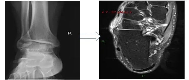
Figure 1: Radiograph and MRI findings of calcaneocuboid coalition
Tarsal coalition is an abnormal connection/ union between two bones, causing non-specific pain in the mid or hind foot due to loss of motion of the affected joint. The condition is more prevalent in males and presents in childhood/early adolescence. The true prevalence of tarsal coalition cannot be determined, as most patients are asymptomatic. However, in comparison to other variants of the foot, tarsal coalition is relatively infrequent.[3]
Discussion
The pathophysiology of tarsal coalition is due to faulty segmentation during development, with resultant abnormal communication seen between bones and a lack of normal joint formation. Calcaneocuboid and talocalcaneal coalitions are the most common. Imaging modalities for diagnosing tarsal coalitions include radiographs, CT, or MRI, with CT being the investigation of choice if radiographs are inconclusive.[4]
The coalition between the calcaneum and the cuboid is very rare, and we could find only 1 case report of this coalition so far. It occurs more often in association with other anomalies of the foot rather than in isolation. It is an important roentgenographic feature of type I acrocephalosyndactyly (Apert’s syndrome). Craig reported a case of Crouzon’s syndrome (a craniofacial dysostosis) which showed associated bilateral isolated calcaneocuboid synostosis.[5] Radiographic findings of tarsal coalition depend upon the tissue bridging between bones, with a large bony bar seen in the case of osseous coalition, whereas in fibrous coalitions, there is irregularity of bony interface, subchondral changes, and even joint space loss. Often, CT and MRI are used due to the lower sensitivity of radiography. MRI can help to distinguish between types of coalition.[6]
Conservative therapy is usually preferred for the early stages of the disease. However, if conservative treatment fails, then surgical excision gives better results, preventing later degeneration of the joint.[7]
Conclusion
Knowledge about the normal anatomy of the foot, particularly the hind foot, combined with varied histological subtypes of tarsal coalition is significant in interpreting radiographs, CT and MRI images and helps in explaining the cause of rigid painful flatfoot in some cases not attributed to other causes.
References
- Linklater J, Hayter CL, Vu D, Tse K. Anatomy of the subtalar joint and imaging of talo-calcaneal coalition. Skeletal Radiol. 2009;38(5):437-449. doi:10.1007/s00256-008-0615-4 PubMed | Crossref | Google Scholar
- Lui TH. Arthroscopic resection of the calcaneonavicular coalition or the “too long” anterior process of the calcaneus. Arthroscopy. 2006;22(8):903.e1-903.e4. doi:10.1016/j.arthro.2005.12.059 PubMed | Crossref | Google Scholar
- Vosoughi AR, Matz J, Rammelt S. Tarsal coalitions: Focusing on calcaneonavicular and talocalcaneal coalitions. Fuß Sprunggelenk. 2023;21(2):150-161. doi:10.1016/j.fuspru.2023.03.002
Tarsal coalitions: Focusing on calcaneonavicular and talocalcaneal coalitions - Worsham J, Neal K, Hahn G, Moran E, Klassen C. Cuboid-navicular coalition in pediatrics: A systematic review and report. Curr Orthop Pract. 2016;27(2):206-211. doi:10.1097/bco.0000000000000353
Cuboid-navicular coalition in pediatrics: A systematic review and report - Imai K, Ikoma K, Kido M, et al. Nonosseous tarsal coalition of the lateral cuneocuboid joint: A case report. J Foot Ankle Surg. 2016;55(5):1072-1075. doi:10.1053/j.jfas.2015.07.008 PubMed | Crossref | Google Scholar
- Efstathopoulos N, Nikolaou V, Lazarettos J, et al. Calcaneonavicular coalition. Eur J Orthop Surg Traumatol. 2006;16:70–74. doi:10.1007/s00590-005-0023-6
Calcaneonavicular coalition
Acknowledgments
Not reported
Funding
Not reported
Author Information
Corresponding Author:
Namrah Khalid
Department of Diagnostic Radiology
Shifa International Hospital Islamabad, Pakistan
Email: namrahk8@gmail.com
Co-Authors:
Mariam Shah, Rashed Nazeer, Samina Akhtar
Department of Diagnostic Radiology
Shifa International Hospital Islamabad, Pakistan
Authors Contributions
Mariam Shah was responsible for the concept, literature review, manuscript writing, and critical review. Namrah Khalid contributed to the concept, literature review, and manuscript writing. Rashed Nazeer handled the analysis and interpretation of the data and performed a critical review. Samina Akhtar contributed to the literature review and critical review.
Informed Consent
Informed consent was obtained from the patient for publication of this case report.
Conflict of Interest Statement
The author declares no conflicts of interest.
Guarantor
None
DOI
Cite this Article
Shah M, Khalid N, Nazeer R, Akhtar S. An Unusual Cause of Painful Foot: Calcaneocuboid Synostosis. medtigo J Med 2025;3(2):e3062324. doi:10.63096/medtigo3062324 Crossref


