


 Author Affiliations
Abstract
An esophageal inlet patch (also known as a gastric inlet patch) is an ectopic gastric mucosa, characterized by salmon-colored mucosa, typically found in the proximal esophagus just below the upper esophageal sphincter. While most cases are asymptomatic, some patients may present symptoms such as dysphagia, globus sensation, cough, and, occasionally, pain. Complications can include stricture, webs, rings, metaplasia, adenocarcinoma, and ulceration with upper gastrointestinal (GI) bleeding. Here, we present the case of a 52-year-old patient with an esophageal inlet patch experiencing typical symptoms.
Keywords
Dysphagia, Heterotopic mucosa, Globus sensation, Endoscopy, Ethiopia.
Introduction
Ectopic gastric mucosa can occur anywhere along the GI tract. When it occurs in the upper esophagus, it is referred to as an “inlet patch (IP)” due to its location at or just distal to the upper esophageal sphincter.[1] Heterotopic gastric mucosa has also been reported in other places including the rectum and the anus as well as the duodenum, jejunum, gallbladder, cystic duct, and ampulla of vater. The etiology and pathological characteristics of these conditions remain unexplored and unclear.[2]
Case presentation
This is a 52-year-old female patient from Gondar who presented with difficulty swallowing both solid and liquid foods, as well as a foreign body sensation in the throat, which has been present for 1 month. She denies any history of weight loss, epigastric pain, or chest pain, and there is no family history of gastrointestinal malignancies. The patient had no known chronic medical illness. On physical examination, blood pressure (BP)-125/90 mmHg, respiratory rate (RR)-18 breaths per minute, pulse rate (PR) 104 bpm, and temperature 36.9°C. She had pink conjunctiva and non-icteric sclera. Examinations in other systems were unremarkable.
Laboratory tests showed normal complete blood count (CBC), serum creatinine, and liver enzymes. The stool Helicobacter pylori (H. pylori) antigen test was negative. Upper GI endoscopy revealed salmon-colored mucosa approximately 17 cm from the incisors in the proximal esophagus (Figure 1). She started on pantoprazole 40 mg per oral (PO) daily and was scheduled for the next visit after 1 month. There was a significant improvement in symptoms during her follow-up visit.
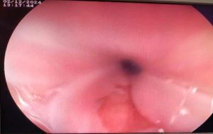
Figure 1: Salmon-colored mucosa at the proximal esophagus (17cm from the incisors)
Discussion
IP is a distinct region of heterotopic gastric mucosa occurring in the proximal one-third of the esophagus, with a prevalence of 0.1% to 10% in patients undergoing upper endoscopy. This wide range in prevalence is due to the special interest of some endoscopists who specifically look for it.[3] The most held theory concerning the origin of the gastric inlet patch (GIP) is the misplacement or sequestration of endoderm from the gastric anlage in the developing esophagus. This process is thought to occur at the 4-week embryonic stage when the primitive stomach lies in the neck region.[4]
Understanding of the clinical significance of IP among patients with pharyngoesophageal complaints continues to evolve. Secretions from IP lesions, such as pepsin and hydrochloric acid, may produce symptoms of laryngopharyngeal reflux (LPR) because of the proximal location of the IP. LPR is defined as reflux above the level of the upper esophageal sphincter, resulting in cough, hoarseness, dysphagia, and/or globus sensation. In a prospective cohort study of 65 patients with IP, the majority experienced heartburn or acid regurgitation (51%), followed by dysphagia (17%), cough (14%), and globus sensation (6%).[5] Mostly, this lesion is asymptomatic. However, it may be associated with structural abnormalities (webs, strictures, ulcers, fistula), causing local symptoms (pain, dysphagia, hoarseness) and rarely, adenocarcinoma. Moreover, there is an association between this condition and Barrett’s esophagus. Although H. pylori may be detected in an IP, ulceration and bleeding are rare.[7]
Patients with no symptoms do not need treatment for a GIP. However, if patients are symptomatic, then first-line medical treatment would be with a proton pump inhibitor (PPI), particularly as the majority of symptoms in these patients are secondary to acid production from the GIP. In patients not responsive to PPI treatment, endoscopic treatment is a potential option. In benign inlet patches, ablative treatment with argon plasma coagulation (APC) or radiofrequency ablation (RFA) is beneficial.[7]
Conclusion
Endoscopic evaluation of the proximal esophagus can be challenging without appropriate sedation. An esophageal IP should be considered in the differential diagnosis of benign causes of dysphagia and globus sensation. Proper management and follow-up are essential to prevent potential complications.
References
- Behrens C, Yen PPW. Esophageal inlet patch. Radiol Res Pract. 2011;2011:460890. doi:10.1155/2011/460890
PubMed | Crossref | Google Scholar - Ciocalteu A, Popa P, Ionescu M, Gheonea DI. Issues and controversies in esophageal inlet patch. World J Gastroenterol. 2019;25(30):4061-4073. doi:10.3748/wjg.v25.i30.4061 PubMed | Crossref | Google Scholar
- Jamma S, Tangella K, Ramkumar D. Cervical inlet patch of the esophagus: a cause for concern? Gastrointest Endosc. 2007;65(5):AB330. doi:10.1016/j.gie.2007.03.230 Crossref | Google Scholar
- Meining A, Bajbouj M. Gastric inlet patches in the cervical esophagus: what they are, what they cause, and how they can be treated. Gastrointest Endosc. 2016;84(6):1027-1029. doi:10.1016/j.gie.2016.08.012
PubMed | Crossref | Google Scholar - Iyer N, Afshar K, Joshua J, et al. Esophageal inlet patch: a clinically significant entity in advanced pulmonary disease and lung transplant rejection. Foregut. 2023;3:263451612211319. doi:10.1177/26345161221131985 Crossref | Google Scholar
- Bataller R, Bordas JM, Ordi J, Llach J, Elizalde JI, Mondelo F. Upper gastrointestinal bleeding: a complication of “inlet patch mucosa” in the upper esophagus. Endoscopy. 1995;27(3):282. doi:10.1055/s-2007-1005690
PubMed | Crossref | Google Scholar - Hussein M, Dunn J. Gastric inlet patch: an under-diagnosed cause of globus. ENT & Audiology News. 2023. Gastric inlet patch: an under-diagnosed cause of globus
Acknowledgments
Not reported
Funding
Not reported
Author Information
Zelalem Mulu
Department of Internal Medicine
Debre Markos University, Debre Markos, Ethiopia
Email: zmlaew@gmail.com
Author Contribution
The author contributed to the conceptualization, investigation, and data curation by acquiring and critically reviewing the selected articles and was involved in the writing – original draft preparation, and writing – review & editing to refine the manuscript.
Ethical Approval
Informed consent was acquired from the patient. The case report was approved by the Research and Ethical Committee of the Hospital.
Conflict of Interest Statement
The author declares no conflict of interest.
Guarantor
None
DOI
Cite this Article
Zelalem M. A Rare Diagnosis in a Common Symptom: Esophageal Inlet Patch. medtigo J Emerg Med. 2025;2(1):e3092215. doi:10.63096/medtigo3092215 Crossref


